the Creative Commons Attribution 4.0 License.
the Creative Commons Attribution 4.0 License.
Additional new organic-walled dinoflagellate cysts from two onshore UK Chalk boreholes
Martin A. Pearce
Beautifully preserved dinoflagellate cysts continue to be discovered in UK Cretaceous chalks and provide important new biostratigraphic information. Five new species – Conosphaeridium norfolkense sp. nov., Glaphyrocysta coniacia sp. nov., Impletosphaeridium banterwickense sp. nov., Sentusidinium devonense sp. nov., Sentusidinium spinosum sp. nov. and the new subspecies Spiniferites ramosus subsp. ginakrogiae subsp. nov. – are described from Upper Cretaceous strata of the British Geological Survey (BGS) Banterwick Barn and Trunch boreholes (onshore UK). An emended diagnosis for Odontochitina diducta Pearce is also provided to broaden the morphological variability in the type material.
- Article
(12573 KB) - Full-text XML
- BibTeX
- EndNote
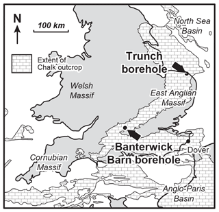
Cretaceous chalks from onshore UK boreholes have recently yielded beautifully preserved dinoflagellate cysts (Prince et al., 1999, 2008; Pearce et al., 2003, 2011; Pearce, 2010) but many undescribed forms continue to be found. The sample material is exceptional in its δ13C chemostratigraphic correlation to outcrop sections with macrobiostratigraphic age control (see Jarvis et al., 2006). This paper describes five new species and one new subspecies from the British Geological Survey (BGS) Banterwick Barn (Berkshire; northern Anglo-Paris Basin) and BGS Trunch borehole (Norfolk, southern North Sea Basin; Fig. 1).
The Banterwick Barn borehole (Berkshire; UK national grid reference SU 5134 7750; 51∘29′39 N, 1∘15′43 W) was cored in 1996 by the BGS as part of a Chalk aquifer study yielding ∼ 97 m of Upper and Middle Chalk. Jarvis et al. (2006) used δ13C isotope stratigraphy to determine an age range of Lower Turonian to Middle Coniacian. Pearce et al. (2003) demonstrated a significant attenuation/unconformity spanning much of the Middle and Upper Turonian. The Trunch borehole (Norfolk; UK national grid reference TG 2933 3455; 52∘51′34 N, 01∘24′19 E), was continuously cored in 1975 by the BGS (then the Institute of Geological Sciences) to sample the Chalk at its most complete development in Britain. The 10 in. (25.4 cm) diameter core recovered a thick Quaternary cover and 468 m of Cenomanian–lower Maastrichtian Chalk (Wood et al., 1994), including 246 m of Campanian strata. The chalk samples from the borehole were taken from composite bags of 10 cm intervals, no cut round of core was preserved. Palynological processing techniques follow that of Pearce et al. (2003). All slides are lodged at the British Geological Survey, Kingsley Dunham Centre, Keyworth, Nottingham, UK. England Finder (EF) coordinates are provided for type and reference specimens. Please note that the slide label must be placed on the right-hand side.
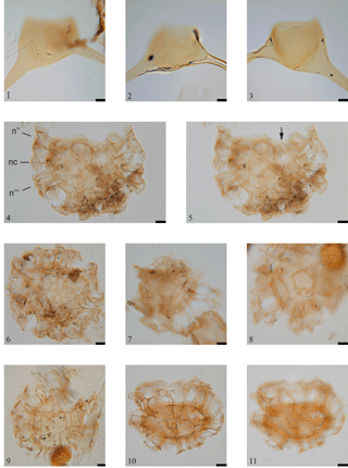
Plate 1(1–3) Odontochitina diducta Pearce 2010, emend. nov.: (1) showing a very narrow hypopericoel connecting the lateral and antapical horns; (2–3) separate cornucave specimens. (4–5) Glaphyrocysta coniacia sp. nov. (holotype), MPK 14626, EF coordinates: M40: (4) internal (reversed) dorsal view, showing a precingular arcuate complex – n, nc and n indicate the precingular, cingular and postcingular series, respectively; (5) external ventral view, with the arrow pointing to the offset sulcal notch. (6) Glaphyrocysta coniacia sp. nov. (paratype 1), MPK 14627, EF coordinates: K46/1: internal (reversed) ventral? view of a complete specimen showing the widely variable process widths. (7) Glaphyrocysta coniacia sp. nov. (paratype 2), MPK 14628, EF coordinates: Q21/2: external view showing the fenestrated distal platforms. (8) Glaphyrocysta coniacia sp. nov. (paratype 3), MPK 14629, EF coordinates: K19: external view showing a dorsal? annulate process. (9) Glaphyrocysta coniacia sp. nov. (paratype 4), MPK 14630, EF coordinates: J11: external dorsal view, showing a partially attached operculum. (10–11) Glaphyrocysta coniacia sp. nov. (paratype 5), MPK 14631, EF coordinates: L22/1: external antapical view showing the dorso-ventral compression. Scale bar represents 10 µm.
Division Dinoflagellata (Bütschli, 1885) Fensome et al., 1993.
Subdivision Dinokaryota Fensome et al., 1993.
Class Dinophyceae Pascher, 1914.
Subclass Peridiniphycidae Fensome et al., 1993.
Order Gonyaulacales Taylor, 1980.
Suborder Ceratiineae Fensome et al., 1993.
Family Ceratiaceae Willey and Hickson, 1909.
Genus Odontochitina Deflandre, 1937.
Type. Odontochitina operculata (Wetzel, 1933)
Deflandre and Cookson, 1955.
Odontochitina diducta Pearce, 2010 emend. nov.
(Pl. 1, figs. 1–3)
1967 Odontochitina costata Alberti, 1961; Clarke and Verdier: pl. 13, fig. 4 only.
1968 Odontochitina sp. Cookson and Eisenack: 112, pl. 2, fig. D.
1991 Odontochitina operculata (Wetzel, 1933) Deflandre and Cookson, 1955; Heine: pl. 27, fig. 15.
1992 Odontochitina sp. Costa and Davey: pl. 3.13, fig. 5.
1997 Odontochitina operculata (Wetzel,
1933) Deflandre and Cookson, 1955; Roncaglia and Corradini: pl. 2,
fig. 5.
Emended diagnosis. A cornucavate to hypocavate species of Odontochitina
with a widely divergent antapical and lateral horn separated by an angle
equal to or greater than 80∘.
Emended description. Large ceratioid, cavate
dinoflagellate cyst with one apical, antapical and lateral horn of comparable
length. The wall is two-layered, comprising a smooth endophragm and smooth,
incompletely and faintly striate or distally perforate (although this may be
due to corrosion) periphragm. The periphragm and endophragm are appressed in
the precingular region with the lateral and antapical horns being either
cornucavate or connected by a hypocavation. The endocyst is sub-spherical,
lacking obvious projections into the pericoel. The antapical and lateral
horns are separated by an angle of greater than 80∘. The paracingulum
may be indicated by faint ridges on the periphragm. The archaeopyle is apical
Type (tA) and the operculum is detached.
Remarks. In the original description of O. diducta, Pearce (2010:62) described the species as having “a well-developed
cavation connecting the lateral and antapical horns”. However, this feature seems to be gradational to specimens from the same material that are cornucavate, and the diagnosis and description are emended here to account for
this fact. The specimens referred to as Odontochitina sp. by Cookson
and Eisenack (1968) and Costa and Davey (1992) are thus now considered
synonymous with O. diducta.
Suborder Gonyaulacineae (autonym).
Family Areoligeraceae Evitt, 1963.
Genus Glaphyrocysta Stover and Evitt, 1978.
Type. Glaphyrocysta retiintexta (Cookson, 1965) Stover and
Evitt, 1978.
Glaphyrocysta coniacia sp. nov.
(Pl. 1, figs. 4–11)
2003 Glaphyrocysta sp. A Pearce et al.: pl. 1,
fig. 8.
Derivation of name. Named after the Coniacian Stage from
which the type material was obtained.
Diagnosis. A species of Glaphyrocysta with a
microreticulate endophragm and a weakly fibrous periphragm forming processes
of highly variable width, united into linear, arcuate, soleate or annulate
complexes that unite distally into wide, perforate, recurved and irregular
membranes.
Holotype. MPK 14626, EF coordinates: M40, Pl. 1,
figs. 4–5.
Paratypes. Paratype 1, MPK 14627, EF coordinates: K46/1,
Pl. 1, fig. 6; paratype 2, MPK 14628, EF coordinates: Q21/2, Pl. 1, fig. 7;
paratype 3, MPK 14629, EF coordinates: K19, Pl. 1, fig. 8; paratype 4, MPK
14630, EF coordinates: J11, Pl. 1, fig. 9; paratype 5, MPK 14631, EF
coordinates: L22/1, Pl. 1, figs. 10–11.
Type locality and horizon. BGS Banterwick Barn
borehole, Berkshire; 1.48–1.55 m, Upper Chalk Formation (Broadstairs Chalk
Member), Micraster coranguinum Zone (Middle Coniacian).
Description. Medium to large chorate dinoflagellate cyst
with a lenticular, dorso-ventrally compressed, central body and an offset
sulcal notch. Two weak antapical bulges may be present. Wall two-layered,
comprising a micro-reticulate endophragm and a slightly fibrous periphragm
that separate in the formation of processes. The processes are tabular,
apparently solid, and are well developed around the ambitus of the central
body, poorly developed on the mid-dorsal area and typically absent on the
mid-ventral area. The marginal processes are of variable width but similar
length on individual specimens and appear to be clustered into linear groups
that are connected distally and may be connected at the base. The mid-dorsal
processes may develop into shorter arcuate, soleate or annulate complexes.
Distally, the processes widen into perforated platforms that are irregular in
shape and typically recurved. The sulcal notch is offset to the left; the
cingulum is represented by slender processes at margins of the central body.
The archaeopyle is apical, Type tA, typically with a detached operculum.
Dimensions. Holotype, central body
w∕l = 74 × 61 µm, maximum process
length = 35 µm; paratype 1, central body
w∕l = 71 × 62 µm, maximum process
length = 27 µm; paratype 2, central body
w∕l = 59 × 53 µm, maximum process
length = 30 µm; paratype 3, central body
w∕l = 60 × 60 µm, maximum process
length = 30 µm; paratype 4, central body
w∕l = 70 × 53 µm, maximum process
length = 25 µm; paratype 5, maximum process
length = 36 µm. Range, central body
w∕l = 46(60)74 × 42(55)64 µm, maximum process
length = 12(29)36 µm. Sixteen specimens measured.
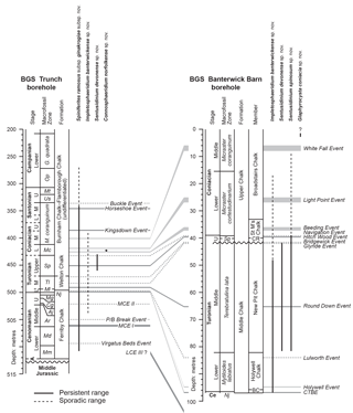
Figure 2Stratigraphic distribution of the new species from the BGS Banterwick Barn and Trunch boreholes, calibrated by the isotopic events of Jarvis et al. (2006). Abbreviations: A, Albian; Aj, Acanthoceras jukesbrownei; BC, Ballard Cliff Member; Ce, Cenomanian; Cg, Calycoceras guerangeri; CTBE, Cenomanian–Turonian Boundary Event; CR, Chalk Rock; G. quadrata, Gonioteuthis quadrata; L, lower; LCE III, Lower Cenomanian Event III; M, middle; M. coranguinum, Micraster coranguinum; Mc, Micraster cortestudinarium; MCE I, Middle Cenomanian Event I; MCE II, Middle Cenomanian Event II; Md, Mantelliceras dixoni; Mg, Metoicoceras geslinianum; Ml, Mytiloides labiatus; Mm, Mantelliceras mantelli; Mt, Marsupites testudinarius; Nj, Neocardioceras juddii; Op, Offaster pilula; Sp, Sternotaxis plana; St M's Chalk, St Margaret's Chalk; Tl, Terebratulina lata; U, upper; Us, Uintacrinus socialis. Modified from Jarvis et al. (2006).
Stratigraphic range. Upper Chalk Formation, Broadstairs
Chalk Member, Micraster coranguinum Zone (Middle Coniacian), above
the White Fall Event in the Banterwick Barn borehole to an unconfirmed upper
limit (Fig. 2).
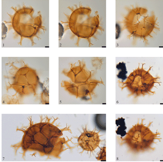
Plate 2(1–3) Spiniferites ramosus subsp. ginakrogiae subsp. nov. (holotype), MPK 14639, EF coordinates: T46: (1) internal (reversed) dorsal view; (2) ambital view; (3) external ventral view. (4) Spiniferites ramosus subsp. ginakrogiae subsp. nov. (paratype 1), MPK 14640, EF coordinates: V19/2: external (right) lateral view. (5) Spiniferites ramosus subsp. ginakrogiae subsp. nov. (paratype 2), MPK 14641, EF coordinates: V26/4: external (left) lateral view. (6, 8) Spiniferites ramosus subsp. ginakrogiae subsp. nov., specimen from the Gina Krog Field, Norwegian North Sea; (7) Spiniferites ramosus subsp. ginakrogiae subsp. nov. (left) and S. ramosus subsp. ramosus (right), size comparison. Scale bar represents 10 µm.
Comparison. The closest species to Glaphyrocysta coniacia sp. nov. are G. exuberans (Deflandre and Cookson, 1955 ex
Eaton, 1976) Stover and Evitt, 1978, G. intricata (Eaton, 1971) Stover
and Evitt, 1978 and G. texta (Bujak, 1976) Stover and Evitt, 1978.
Despite being described from the Palaeogene and therefore being significantly
younger, Glaphyrocysta exuberans differs with processes
that ramify elaborately halfway along their length, while G. intricata and G. texta show much less variability in process width.
Glaphyrocysta intricata also differs in having processes that
bifurcate distally to a variable length and width.
Family Gonyaulacaceae Lindemann, 1928.
Subfamily Gonyaulacoideae (autonym).
Genus Spiniferites Mantell, 1850.
Type. Spiniferites ramosus (Ehrenberg, 1837) Mantell, 1854.
Spiniferites ramosus subsp. ginakrogiae subsp. nov.
(Pl. 2, figs. 1–6, 7 (left), 8)
Derivation of name. After the Norwegian Gina Krog and oil
field (in the Norwegian North Sea) of the same name, where the species has
also been recognised.
Diagnosis. A large subspecies of Spiniferites ramosus with a smooth wall, narrow sutural crests and intergonal processes
on some of the larger plate boundaries.
Holotype. MPK 14639, EF coordinates: T46, Pl. 2,
figs. 1–3.
Paratypes. Paratype 1, MPK 14640, EF coordinates: V19/2,
Pl. 2, fig. 4; paratype 2, MPK 14641, EF coordinates: V26/4, Pl. 2, fig. 3.
Type locality and horizon. BGS Trunch borehole, Norfolk;
451.0 m, Upper Chalk (Burnham Chalk Formation), Sternotaxis
plana Zone (Upper Turonian).
Description. Large, spiniferate, chorate dinoflagellate
cyst with a sub-spherical central body. The wall is two-layered with a thick
(∼ 2 µm) and smooth endophragm and a thin
(∼ 0.5 µm) periphragm, the latter of which develops solid
processes. Distally trifurcating gonal and bifurcating intergonal processes
(on the boundaries between the larger precingular, cingular and postcingular
plates) are typically less than one-third the central body diameter, with the
furcation occurring from 50 % (on shorter processes) to 25 % (on
longer processes) from the distal end. Narrow sutural crests line the
processes, often to the distal extremity, occasionally rendering the bi- and
trifurcations relatively wide, and define a clear standard gonyaulacacean
tabulation. The cingulum is weakly laevorotatory, typically by one cingulum
width; the parasulcus lacks clearly developed sutures. The archaeopyle is
precingular, Type P, operculum detached.
Dimensions. Holotype: central body w∕l:
74 × 75 µm, maximum process length = 22 µm,
paratype 1: central body w∕l: 71 × 76 µm, maximum process
length = 29 µm, paratype 2: central body w∕l:
91 × 80 µm, maximum process length = 19 µm.
Range central body
w∕l = 54(69)91 µm × 50(69)84 µm, maximum
process length, 15(21)30 µm. Twenty specimens measured.
Stratigraphic range. Ferriby Chalk Formation,
Mantelliceras mantellii Zone (Lower Cenomanian; questionably between
the Lower Cenomanian Event I and the Virgatus Beds Event) to the
Burnham–Flamborough Chalk Formation (undifferentiated), Gonioteuthis quadrata Zone (Lower Campanian) in the Trunch borehole (Fig. 2).
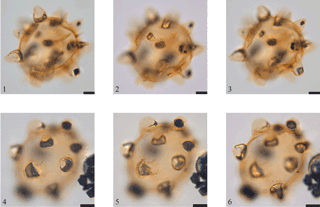
Plate 3(1–3) Conosphaeridium norfolkense sp. nov. (holotype), MPK 14624, EF coordinates: K37/1: internal (reversed) view of the precingular archaeopyle; precise orientation not determined. (4–6) Conosphaeridium norfolkense sp. nov. (paratype), MPK 14625, EF coordinates: V26/1: external view; precise orientation not determined. Scale bar represents 10 µm.
Remarks. This species appears to be morphologically intermediate between Spiniferites ramosus subsp. gracilis (by possessing intergonal processes) and Spiniferites porosus (in size). The central body diameter of Spiniferites porosus ranges from 66 to 69 µm (the holotype is 66 × 75 µm), with process lengths ranging from 17 to 36 µm (holotype 17–23 µm) long.
Comparison. Differs from other subspecies of
Spiniferites ramosus by its conspicuously large size.
Subfamily Leptodinioideae Fensome et al., 1993.
Genus Conosphaeridium Cookson and Eisenack, 1969.
Type. Conosphaeridium striatoconum (Deflandre and
Cookson, 1955) Cookson and Eisenack, 1969.
Conosphaeridium norfolkense sp. nov.
(Pl. 3, figs. 1–6)
1976 Conosphaeridium cf. striatoconum of
Benson: pl. 1, figs. 4–5.
Derivation of name. Named after the English county of
Norfolk, from where the type material was obtained.
Diagnosis. A species of Conosphaeridium with a
smooth to only very weakly striate periphragm.
Holotype. MPK 14624, EF coordinates: K37/1, Pl. 3,
figs. 1–3.
Paratype. MPK 14625, EF coordinates: V26/1, Pl. 3,
figs. 4–6.
Type locality and horizon. BGS Trunch borehole, Norfolk;
423.0 m, Upper Chalk (Burnham Chalk Formation), Micraster
cortestudinarium Zone (Lower Coniacian).
Description. Medium-sized proximochorate dinoflagellate
with a globular sub-spherical central body. The wall is two-layered,
comprised of a thin, smooth endophragm and smooth to very weakly striate
periphragm that forms hollow intratabular processes. The processes are
relatively squat, rounded-lagenate and typically open distally. The
archaeopyle is precingular, Type P, the operculum may be attached,
but more usually detached.
Dimensions. Holotype, central body
diameter = 60 µm, maximum processes
length = 17 µm. Paratype, central body
diameter = 51 µm, maximum processes
length = 12 µm. Range, central body
diameter = 44(54)65 µm, maximum processes
length = 8(12)17 µm. Six specimens measured.
Stratigraphic range. Only observed from the sample
containing the holotype, between the Beeding and Light Point events (Fig. 2).
Comparison. Both Conosphaeridium
abbreviatum Wilson, 1984 and C. striatoconum (Deflandre and
Cookson, 1955) Cookson and Eisenack, 1969 differ by possessing strong ribs on
the processes; the former also has a much larger central body size (holotype
body diameter 95 × 81 µm). Brideaux and McIntyre (1975,
pl. 7, figs. 17–18) figured a specimen as Conosphaeridium sp.
A from the Middle Albian of northern Canada that is similar to C. norfolkense in possessing a very weakly striate periphragm, but which
differs in the shape of the processes that are tubular and narrow distally
with a truncated distal margin. The specimen figured as
Conosphaeridium cf. striatoconus by Benson (1976) from the
undifferentiated Upper Cretaceous of Maryland, USA, is considered here
synonymous with C. norfolkense sp. nov.
Subfamily uncertain
Genus Sentusidinium Sarjeant and Stover, 1978.
Type. Sentusidinium rioultii (Sarjeant, 1968) Sarjeant and
Stover, 1978.
Remarks. As stated by Wood et al. (2016) in their
emendation of the genus, the operculum of Sentusidinium is normally
detached (implying that attached operculae are permitted), and deep accessory
archaeopyle sutures are typically present.
Sentusidinium devonense sp. nov.
(Pl. 4, figs. 1–12)
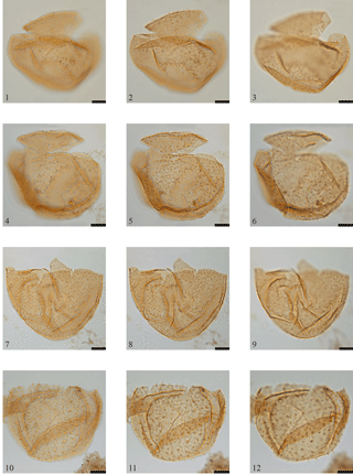
Plate 4(1–3) Sentusidinium devonense sp. nov. (holotype), MPK 14634, EF coordinates: O17: (1) external view; (3) internal (reversed) view. (4–6) Sentusidinium devonense sp. nov. (paratype 1), MPK 14635, EF coordinates: F37/3: (4) external view; (6) internal (reversed) view. (7–9) Sentusidinium devonense sp. nov. (paratype 2), MPK 14636, EF coordinates: D50/4: (7) external view; (9) internal (reversed) view. (10–12) Sentusidinium devonense sp. nov. (paratype 3), MPK 14637, EF coordinates: L16: (10) external view; (12) internal (reversed) view. Scale bar represents 10 µm.
1987 Sentusidinium sp. B in Tocher and Jarvis: pl. 9.3, figs. 7–9.
1995 Canningia sp. B FitzPatrick: fig. 9n.
Derivation of name. From the English county of Devon, from
where the species was originally recorded.
Diagnosis. A species of Sentusidinium with an even
covering of short, flexuous and acuminate spines.
Holotype. MPK 14634, EF coordinates: O17, Pl. 4,
figs. 1–3.
Paratypes. Paratype 1, MPK 14635, EF coordinates: F37/3,
Pl. 4, figs. 4–6; paratype 2, MPK 14636, EF coordinates: D50/4, Pl. 4,
figs. 7–9; paratype 3, MPK 14637, EF coordinates: L16, Pl. 4, figs. 10–12.
Type locality and horizon. BGS Banterwick Barn borehole,
Berkshire; 48.06–48.09 m, Middle Chalk Formation, New Pit Chalk Member,
Terebratulina lata Zone (Middle Coniacian).
Description. A medium-sized proximate dinoflagellate cyst
with a sub-rounded body. The wall is composed of a finely granular autophragm
that possesses short (∼ 2 µm), evenly distributed and
apparently solid, simple flexuous spines with acuminate tips. No expression
of the cingulum or sulcus is present. The archaeopyle is apical, Type tA,
with a zig-zag margin and clear accessory sutures. The operculum may be
attached but more usually detached.
Dimensions. Holotype, central body w∕l (excluding
operculum) = 66 × 43 µm, maximum process length
2 µm; paratype 1, central body w∕l (excluding
operculum) = 65 × 48 µm, maximum process length
2 µm; paratype 2, central body
w∕l = 68 × 50 µm, maximum process length
2 µm; paratype 3, central body
w∕l = 60 × 50 µm, maximum process length
3 µm. Range, central body w∕l (excluding
operculum) = 40(61)76 × 40(48)60 µm, maximum process
length = 2(2)3 µm. Seventeen specimens measured.
Stratigraphic range. Middle Chalk Formation, Holywell Chalk
Member, Mytiloides labiatus/Terebratulina
lata boundary (Middle Turonian; between the Lulworth and Round Down
events in the Banterwick Barn borehole) to the Burnham–Flamborough Chalk
Formation (undifferentiated), Micraster cortestudinarium
Zone (Lower Coniacian; between the Navigation and Beeding events in the
Trunch borehole; Fig. 2).
Remarks. FitzPatrick (1995) described the informal species
Canningia sp. B from the Turonian of Southern England. She
considered the generic attribution based on the subspherical outline,
non-tabular spinose ornament and apical archaeopyle; however, she also
mentioned the lack of an offset sulcal notch. These observations suggest that
the species is not an areoligeracean, and that it has a greater affinity with
Sentusidinium, and it is here considered synonymous with S.
devonense sp. nov.
Comparison. For such a simple genus of dinoflagellate cyst,
there are a number of comparable species, but which all differ in the shape
of the central body and/or the morphology of the processes.
Sentusidinium aptiense (Burger, 1980a) Burger, 1980b possesses hollow
tubular spines and S. capillatum (Davey, 1975) Lentin and Williams,
1981, S. echinatum (Gitmez and Sarjeant, 1972) Sarjeant and Stover,
1978 and S? millepiedii (Jain and Millepied,
1975) Islam, 1993 possesses much more densely distributed ornament.
Sentusidinium capitatum (Cookson and Eisenack, 1960) Wood et al.,
2016 may possess short spines with acuminate tips but the body is distinctly
elongate, while S. minus (Jiabo, 1978) He et al. in He et al., 1989
is significantly smaller. Sentusidinium myriatrichum Fensome, 1979
possesses significantly shorter and denser ornament and S. perforoconum (Yun Hyesu, 1981) Islam, 1993 has a densely perforate periphragm.
Sentusidinium pilosum (Ehrenberg, 1854) Sarjeant and Stover, 1978 has
a denser cover of short processes with variable tips that are also variable
in S. rioultii Sarjeant, 1968, S. sahii (Khanna and Singh,
1981) Wood et al., 2016, S. sparsibarbatum Erkmen and Sarjeant, 1980
and S. villersense (Sarjeant, 1968) Sarjeant and Stover, 1978.
Sentusidinium separatum (McIntyre and Brideaux, 1980) Lentin and
Williams, 1981 has bifid and branched process tips.
Sentusidinium spinosum sp. nov.
(Pl. 5, figs. 1–6)
Derivation of name. From the Latin spinosum,
meaning spiny.
Diagnosis. A species of Sentusidinium with an even
covering of relatively long, flexuous and acuminate spines.
Holotype. MPK 14638, EF coordinates: S13/4, Pl. 5,
figs. 1–6.
Type locality and horizon. BGS Banterwick Barn borehole,
Berkshire; 39.55–39.59 m, Upper Chalk Formation, St. Margaret's Chalk
Member, Sternotaxis plana Zone (Middle Turonian).
Description. A medium-sized proximate dinoflagellate cyst
with a sub-rounded body. The wall is composed of a micro-reticulate
autophragm that possesses relatively long (4–16 µm), evenly
distributed, hollow spines with simple acuminate tips. No expression of the
cingulum or sulcus is present. The archaeopyle is apical, Type tA, with a
zig-zag margin and clear accessory sutures. The operculum may be attached but
more usually detached.
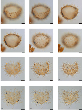
Plate 5(1–6) Sentusidinium spinosum sp. nov. (holotype), MPK 14638, EF coordinates: S13/4: (1) internal (reversed) view, showing the archaeopyle margin; (6) external view of the antapical region. (7–9) Impletosphaeridium banterwickense sp. nov. (holotype), MPK 14632, EF coordinates: Q35/3: (7) external view; (9) internal (reversed) view. (9–12) Impletosphaeridium banterwickense sp. nov. (paratype), MPK 14633, EF coordinates: K6/3: (10) external view; (12) internal (reversed) view. Scale bar represents 10 µm.
Dimensions. Holotype, central body
w∕l = 61 × 48 µm, maximum process
length = 11 µm. Range, central body
w∕l = 46(56)72 × 30(46)56 µm, maximum processes
length = 4(6)16 µm. Twenty specimens measured.
Stratigraphic range. Middle Chalk Formation, Holywell Chalk
Member, Mytiloides labiatus/Terebratulina
lata boundary (Middle Turonian; between the Lulworth and Round Down
events) to the Upper Chalk Formation, Broadstairs Chalk Member,
Micraster coranguinum Zone (Middle Coniacian; below the
White Fall Event) in the Banterwick Barn borehole (Fig. 2).
Comparison. Sentusidinium aptiense
(Burger, 1980a) Burger, 1980b also possesses hollow spines but which differ
from S. spinosum sp. nov. in their relatively shorter length of
3–4 µm. These species also have mutually exclusive ranges, being
Callovian to Aptian for S. aptiense (Wood et al., 2015) and
Turonian for S. spinosum sp. nov. Sentusidinium
densicomatum (Maier, 1959) Sarjeant, 1983 differs in possessing more
densely distributed hair-like projections. The processes are also much more
numerous on S. pilosum (Ehrenberg, 1854) Sarjeant and Stover, 1978
and which also have variable tips. Spine length is comparable on S. sahii (Khanna and Singh, 1981) Wood et al., 2016 and S. seperatum
(McIntyre and Brideaux, 1980) Lentin and Williams, 1981 but which also have
variable tips in the former and bifid and branched tips in the latter.
Suborder uncertain
Family uncertain
Genus Impletosphaeridium Morgenroth, 1966.
Type. Impletosphaeridium transfodum Morgenroth, 1966.
Impletosphaeridium banterwickense sp. nov.
(Pl. 5, figs. 7–12)
Derivation of name. Named after Banterwick Barn, the
borehole from where the species is described.
Diagnosis. A species of Impletosphaeridium
possessing spines that terminate distally into a simple bifurcation with
endings of equal length.
Holotype. MPK 14632, EF coordinates: Q35/2, Pl. 5,
figs. 7–9.
Paratype. MPK
14633, EF coordinates: K6/3, Pl. 5, figs. 10–12.
Type locality and horizon. BGS Banterwick Barn borehole, Berkshire; 79.51–79.53 m, Middle Chalk
Formation, New Pit Chalk Member, Terebratulina lata Zone
(Middle Turonian).
Description. A small chorate dinoflagellate cyst with a
sub-rounded body. The wall is composed of a finely granular autophragm from
which arises evenly distributed, non-tabular solid spines, which terminate in
a short bifurcation. No expression of the cingulum or sulcus is present. The
archaeopyle is apical, Type tA, with a zig-zag margin and accessory sutures.
The operculum may be attached but more usually detached.
Dimensions. Holotype,
central body w∕l = 46 × 39 µm, maximum process
length = 12 µm. Paratype, central body
w∕l = 46 × 39 µm, maximum process
length = 14 µm. Range, central body
w∕l = 35(41)47 × 29(36)43 µm, maximum process
length = 10(13)16 µm. Twenty specimens measured.
Stratigraphic range. Middle
Chalk Formation, Ballard Cliff Member (uppermost Cenomanian; upper
Cenomanian–Turonian Boundary Event) in the Banterwick Barn borehole to the
Burnham–Flamborough Chalk Formation (undifferentiated), Micraster
coranguinum Zone (Middle Coniacian; between the White Fall and
Kingsdown events) in the Trunch borehole (Fig. 2).
Comparison.
Impletosphaeridium furcillatum (Prössl, 1990 ex
Prössl, 1992) Williams et al., 1998 differs in possessing thicker
bifurcate to multifurate processes, while I. ligospinosum (de
Coninck, 1969) Islam, 1983 differs in possessing bifurcations of unequal
length. In I. varispinosum (Sarjeant, 1959) Islam, 1993, the spines
are more numerous and differ in being occasionally simple but more
frequently bifurcate, clavate or “hammer-headed”. The only similar species
of comparable age is I. williamsii (Boltenhagen, 1977) Islam, 1993; although it possesses acuminate to bifurcate processes, many of the latter
style more closely resemble flared process endings than bifurcating spines. Species
of Downiesphaeridium differ in possessing hollow (and distally
closed) processes, the closest species of which is D. aciculare
(Davey, 1969) Islam, 1993, which differs further by possessing wider (although
relatively thin), blade-like processes. According to Davey (1969), the
processes of Downiesphaeridium aciculare are always pointed
distally and occasionally bear small subsidiary spines near their
extremities. In I. banterwickense sp. nov., the subsidiary spines
occur at the extremity and are significantly longer.
All slides are lodged at the British Geological Survey, Kingsley Dunham Centre, Keyworth, Nottingham, UK. England Finder (EF) coordinates are provided for type and reference specimens.
The author declares that he has no conflict of interest.
My thanks to the British Geological Survey for access to the Banterwick Barn
and Trunch borehole material and to Malcolm Jones (Palynological Laboratory
Services) for preparing the Trunch palynological slides. My sincere thanks to
reviewers Chris Clowes and Iain Prince and the editor, Francesca Sangiorgi,
whose comments much improved an earlier draft of the manuscript. Printing
costs were covered by Evolution Applied Limited.
Edited by: Francesca Sangiorgi
Reviewed by:
Chris Clowes and Iain Prince
Alberti, G.: Zur Kenntnis mesozoischer und alttertiärer Dinoflagellaten und Hystrichosphaerideen von Nord- und Mitteldeutschland sowie einigen anderen europäischen Gebieten, Paläeontographica, Abteilung A, 116, 1–58, 1961.
Benson, D. G.: Dinoflagellate taxonomy and biostratigraphy at the Cretaceous-Tertiary boundary, Round Bay, Maryland, Tulane Studies in Geology and Paleontology, 12, 169–233, 1976.
Boltenhagen, E.: Microplancton du Crétacé supérieur du Gabon, Cahiers de paléontologie, un-numbered: 1–150, 1977.
Brideaux, W. W. and McIntyre, D. J.: Miospores and microplankton from Aptian–Albian rocks along Horton River, District of Mackenzie, Geological Survey of Canada, Bulletin, 252, 1–85, 1975.
Bujak, J. P.: An evolutionary series of Late Eocene dinoflagellate cysts from southern England, Mar. Micropaleontol., 1, 101–117, 1976.
Burger, D.: Palynological studies in the Lower Cretaceous of the Surat Basin, Australia. Bureau of Mineral Resources, Geology and Geophysics, Bulletin, 189, 1–106, 1980a.
Burger, D.: Early Cretaceous (Neocomian) microplankton from the Carpentaria Basin, northern Queensland, Alcheringa, 4, 263–279, 1980b.
Bütschli, O.: Erster Band. Protozoa, in: Dr. H.G. Bronn's Klassen und Ordnungen des Thier-Reichs, wissenschaftlich dargestellt in Wort und Bild, C.F. Winter'sche Verlagsbuchhandlung, Leipzig, 865–1088, 1885.
Clarke, R. F. A. and Verdier, J.-P.: An investigation of microplankton assemblages from the Chalk of the Isle of Wight, England, Verhandelingen der Koninklijke Nederlandse Akademie van Wetenschappen, Afdeeling Natuurkunde, Eerste Reeks, 24, 1–96, 1967.
Cookson, I. C.: Cretaceous and Tertiary microplankton from south-eastern Australia, Proceedings of the Royal Society of Victoria, 78, 85–93, 1965.
Cookson, I. C. and Eisenack, A.: Upper Mesozoic microplankton from Australia and New Guinea, Palaeontology, 2, 243–261, 1960.
Cookson, I. C. and Eisenack, A.: Microplankton from two samples from Gingin Brook No.4 borehole, Western Australia, Journal of the Royal Society of Western Australia, 51, 110–122, 1968.
Cookson, I. C. and Eisenack, A.: Some microplankton from two bores at Balcatta, Western Australia, Journal of the Royal Society of Western Australia, 52, 3–8, 1969.
Costa, L. I. and Davey, R. J.: Dinoflagellate cysts of the Cretaceous System, in: A Stratigraphic Index of Dinoflagellate Cysts, edited by: Powell, A. J., Special Publication of the British Micropalaeontological Society, London, 99–131, 1992.
Davey, R. J.: Non-calcareous microplankton from the Cenomanian of England, northern France and North America, part I. British Museum (Natural History) Geology, Bulletin, 17, 103–180, 1969.
Davey, R. J.: A dinoflagellate cyst assemblage from the Late Cretaceous of Ghana. Proceedings of the 5th West African Colloquium on Micropaleontology, 7, 150–173, 1975.
de Coninck, J.: Dinophyceae et Acritarcha de l'Yprésien du sondage de Kallo, Mémoires de l'Institut royal des sciences naturelles de Belgique, 161, 1–67, 1969.
Deflandre, G.: Microfossiles des silex crétacés. Deuxième partie. Flagellés incertae sedis. Hystrichosphaeridés. Sarcodinés. Organismes divers, Ann. Paleontol., 26, 51–103, 1937.
Deflandre, G. and Cookson, I. C.: Fossil microplankton from Australian Late Mesozoic and Tertiary sediments, Aust. J. Mar. Fres. Res., 6, 242–313, 1955.
Eaton, G. L.: A morphogenetic series of dinoflagellate cysts from the Bracklesham Beds of the Isle of Wight, Hampshire, England, in: Proceedings of the 2nd Planktonic Conference, edited by: Farinacci, A., Rome, 1970, 355–379, 1971.
Eaton, G. L.: Dinoflagellate cysts from the Bracklesham Beds (Eocene) of the Isle of Wight, southern England, British Museum (Natural History) Geology, Bulletin, 26, 227–332, 1976.
Ehrenberg, C. G.: Über das Massenverhältniss der jetzt lebenden Kiesel-Infusorien und über ein neues Infusorien-Conglomerat als Polirschiefer von Jastraba in Ungarn. Abhandlungen der Königlichen Akademie der Wissenschaften zu Berlin, aus dem Jahre 1836, Physikalische Klasse, 109–135, 1837.
Ehrenberg, C. G.: Mikrogeologie: das Erden- und Felsen-schaffende Wirken des unsichtbaren kleinen selbständigen Lebens auf der Erde, Leopold Voss, Leipzig, 374+31+88 pp., 1854.
Erkmen, U. and Sarjeant, W. A. S.: Dinoflagellate cysts, acritarchs and tasmanitids from the uppermost Callovian of England and Scotland: with a reconsideration of the “Xanthidium pilosum” problem, Geobios, Lyon, 13, 45–99, 1980.
Evitt, W. R.: A discussion and proposals concerning fossil dinoflagellates, hystrichospheres, and acritarchs, II. National Academy of Sciences, Washington, Proceedings, 49, 298–302, 1963.
Fensome, R. A.: Dinoflagellate cysts and acritarchs from the Middle and Upper Jurassic of Jameson Land, east Greenland, Grønlands Geologiske Undersøgelse, Bulletin, 132, 1–98, 1979.
Fensome, R. A., Taylor, F. J. R., Norris, G., Sarjeant, W. A. S., Wharton, D. I., and Williams, G. L.: A Classification of Living and Fossil Dinoflagellates, Micropaleontological Press, Special Publication, 7, 351 pp., 1993.
FitzPatrick, M. E. J.: Dinoflagellate cyst biostratigraphy of the Turonian (Upper Cretaceous) of southern England, Cretaceous Res., 16, 757–791, 1995.
Gitmez, G. U. and Sarjeant, W. A. S.: Dinoflagellate cysts and acritarchs from the Kimmeridgian (Upper Jurassic) of England, Scotland and France. British Museum (Natural History) Geology, Bulletin, 21, 171–257, 1972.
Guerstein, G. R., Fensome, R. A., and Williams, G. L.: A new areoligeracean dinoflagellate from the Miocene of offshore eastern Canada and its evolutionary implications, Paleontology, 41, 23–34, 1998.
He, C., Shenzhao, Z., and Guangxing, J.: Early Tertiary microphytoplankton from the Dongpu Region. Series on Stratigraphy and Palaeontology of Oil and Gas Bearing Areas in China. Research Institute of Exploration and Development, Zhongyuan Petroleum Exploration Bureau, Nanjing Institute of Geology and Palaeontology, Academia Sinica – The Petroleum Industry Press, Nanjing, China, 99 pp., 1989.
Heine, C. J.: Late Santonian to Early Maastrichtian dinoflagellate cysts of northeast Texas, in: Stratigraphy and micropalaeontology of the Campanian shelf in northeast Texas, edited by: Thompson, L. B., Heine, C. J., Percival, S. F., and Selznick, M. R., 117–147, Micropalaeontology, Special Publication, 5, 1991.
Islam, M. A.: Dinoflagellate cysts from the Eocene Cliff sections of the Isle of Sheppey, Southeast England, Revue de Micropaléontologie, 25, 231–250, 1983.
Islam, M. A.: Review of the fossil dinoflagellate Cleistosphaeridium, Revista española de micropaleontología, 25, 81–94, 1993.
Jain, K. P. and Millepied, P.: Cretaceous microplankton from Senegal Basin, W. Africa, pt. II. Systematics and biostratigraphy, Geophytology, 5, 126–171, 1975.
Jarvis, I., Gale, A. S., Jenkyns, H. C., and Pearce, M. A.: Secular variation in Late Cretaceous carbon isotopes: a new δ13C carbonate reference curve for the Cenomanian–Campanian (99.6–70.6 Ma), Geol. Mag., 143, 561–608, 2006.
Jiabo: On the Paleogene Dinoflagellates and Acritarchs From the Coastal Region of Bohai. Nanjing Institute of Geology and Palaeontology, Academia Sinica, Nanjing, China, 190 pp., 1978.
Khanna, A. K. and Singh, H. P.: Some new dinoflagellates, spores and pollen grains from the Subathu Formation (Upper Palaeocene–Eocene) of Simla Hills, India, Himal. Geol., 9, 385–419, 1981.
Lentin, J. K. and Williams, G. L.: Fossil dinoflagellates: index to genera and species, 1981 Edn., Bedford Institute of Oceanography, Report Series, BI-R-81-12, 345 pp., 1981.
Lindemann, E.: Abteilung Peridineae (Dinoflagellatae), in: Die Natürlichen Pflanzenfamilien nebst ihren Gattungen und wichtigeren Arten insbesondere den Nutzpflanzen, edited by: Engler, A. and Prantl, K., Zweite stark vermehrte und verbesserte Auflage herausgegeben von A. Engler. 2 Band, Wilhelm Engelmann, Leipzig, 3–104, 1928.
Maier, D.: Planktonuntersuchungen in tertiären und quartären marinen Sedimenten, Ein Beitrag zur Systematik, Stratigraphie und Ökologie der Coccolithophorideen, Dinoflagellaten und Hystrichosphaerideen vom Oligozän bis zum Pleistozän, Neues Jahrbuch für Geologie und Paläontologie, Abhandlungen, 107, 278–340, 1959.
Mantell, G. A.: A Pictorial Atlas of Fossil Remains Consisting of Coloured Illustrations Selected from Parkinson's “Organic Remains of a Former World”, and Artis's “Antediluvian Phytology”, Henry G. Bohn, London, UK, xii+207 pp., 1850.
Mantell, G. A.: The Medals of Creation; or, First Lessons in Geology and the Study of Organic Remains, 2nd Edn., 930 pp., 6 pl. (in two volumes); Henry G. Bohn, London, UK, 1854.
McIntyre, D. J. and Brideaux, W. W.: Valanginian miospore and microplankton assemblages from the northern Richardson Mountains, District of Mackenzie, Canada, Geological Survey of Canada, Bulletin, 320, 57 pp., 1980.
Morgenroth, P.: Mikrofossilien und Konkretionen des nordwesteuropäischen Untereozäns, Palaeontographica, Abteilung B, 119, 1–53, 1966.
Pascher, A.: Über Flagellaten und Algen, Deutsche Botanische Gesellschaft, Berichte, 32, 136–160, 1914.
Pearce, M. A.: New organic-walled dinoflagellate cysts from the Cenomanian to Maastrichtian of the Trunch borehole, UK, J. Micropalaeontology, 29, 51–72, 2010.
Pearce, M. A., Jarvis, I., Swan, A. R. H., Murphy, A. M., Tocher, B. A., and Edmunds, W. M.: Integrating palynological and geochemical data in a new approach to palaeoecological studies: Upper Cretaceous of the Banterwick Barn Chalk borehole, Berkshire, UK, Mar. Micropaleontol., 47, 271–306, 2003.
Pearce, M. A., Lignum, J. S., and Jarvis, I.: Senoniasphaera turonica (Prössl, 1990 ex Prössl, 1992) comb. nov., senior synonym of Senoniasphaera rotundata alveolata Pearce et al., 2003: an important dinocyst marker for the Lower Turonian chalk of NW Europe, J. Micropalaeontol., 30, 91–93, 2011.
Prince, I. M., Jarvis, I., and Tocher, B. A.: High-resolution dinoflagellate cyst biostratigraphy of the Santonian–Basal Campanian (Upper Cretaceous): New data from Whitecliff, Isle of Wight, England, Rev. Palaeobot. Palyno., 105, 143–169, 1999.
Prince, I. M., Jarvis, I., Pearce, M. A., and Tocher, B. A.: Dinoflagellate cyst biostratigraphy of the Coniacian–Santonian (Upper Cretaceous): new data from the English Chalk, Rev. Palaeobot. Palyno., 150, 59–96, 2008.
Prössl, K. F.: Dinoflagellaten der Kreide – Unter-Hauterive bis Ober-Turon – im niedersächsischen Becken. Stratigraphie und Fazies in der Kernbohrung Konrad 101 sowie einiger anderer Bohrungen in Nordwestdeutschland, Palaeontographica, Abteilung B, 218, 93–191, 1990.
Prössl, K. F.: Eine Dinoflagellatenpopulation aus dem Eozän von Garoe (Somalia, Ost-Afrika), Giessener Geologische Schriften, 48, 101–123, 1992.
Rawson, P. F.: The Cretaceous, in: Geology of England and Wales, edited by: Duff, P. M. D. and Smith, A. J., Geological Society, London, 355–388, 1992.
Roncaglia, L. and Corradini, D.: Upper Campanian to Maastrichtian dinoflagellate zonation in the northern Apennines, Italy, Newsletters on Stratigraphy, 35, 29–57, 1997.
Sarjeant, W. A. S.: Microplankton from the Cornbrash of Yorkshire, Geol. Mag., 96, 329–346, 1959.
Sarjeant, W. A. S.: Microplankton from the Upper Callovian and Lower Oxfordian of Normandy, Revue de micropaléontologie, 10, 221–242, 1968.
Sarjeant, W. A. S.: A restudy of some dinoflagellate cyst holotypes in the University of Kiel collections. IV. The Oligocene and Miocene holotypes of Dorothea Maier (1959), Meyniana, 35, 85–137, 1983.
Sarjeant, W. A. S. and Stover, L. E.: Cyclonephelium and Tenua: a problem in dinoflagellate cyst taxonomy, Grana, 17, 47–54, 1978.
Stover, L. E. and Evitt, W. R.: Analyses of pre-Pleistocene organic-walled dinoflagellates. Stanford University Publications, Geological Sciences, 15, 300 pp., 1978.
Taylor, F. J. R.: On dinoflagellate evolution, BioSystems, 13, 65–108, 1980.
Tocher, B. A. and Jarvis, I.: Dinoflagellate cysts and stratigraphy of the Turonian (Upper Cretaceous) chalk near Beer, southeast Devon, England, in: Micropalaeontology of Carbonate Environments, edited by: Hart, M. B., Ellis Horwood, British Micropalaeontology Society Series, 138–175, 1987.
Wetzel, O.: Die in organischer Substanz erhaltenen Mikrofossilien des baltischen Kreide-Feuersteins mit einem sedimentpetrographischen und stratigraphischen Anhang, Paläeontographica, Abteilung A, 77, 141–186, 1933.
Willey, A. and Hickson, S. J.: The Protozoa (continued). Section F. – The Mastigophora, in: A Treatise on Zoology. Part 1. Introduction and Protozoa, edited by: Lankester, R., First Fascicle, 154–192, Adam and Charles Black, London (reprinted by A. Asher, Amsterdam, 1964), 1909.
Williams, G. L., Lentin, J. K., and Fensome, R. A.: The Lentin and Williams Index of fossil dinoflagellates 1998 edition. American Association of Stratigraphic Palynologists, Contributions Series, 34, 817 pp., 1998.
Wilson, G. J.: Some new dinoflagellate species from the New Zealand Haumurian and Piripauan stages (Santonian–Maastrichtian, Late Cretaceous), New Zeal. J. Bot., 22, 549–556, 1984.
Wood, C. J., Morter, A. A., and Gallois, R. W.: Appendix 1. Upper Cretaceous stratigraphy of the Trunch borehole. TG23SE8, in: Geology of the Country around Great Yarmouth, edited by: Arthurton, R. S., Booth, S. J., Morigi, A. N., Abbott, M. A. W., and Wood, C. J., Memoir for 1:50,000 Sheet 162 (England and Wales) with an Appendix on the Trunch Borehole by Wood and Morter, HMSO, London, 105–110, 1994.
Wood, S. E. L., Riding, J. B., Fensome, R. A., and Williams, G. L.: A review of the Sentusidinium complex of dinoflagellate cysts, Rev. Palaeobot. Palynol., 234, 61–93, 2016.
Yun, H. S.: Dinoflagellaten aus der Oberkreide (Santon) von Westfalen, Palaeontographica, Abteilung B, 177, 1–89, 1981.


