
the Creative Commons Attribution 4.0 License.
the Creative Commons Attribution 4.0 License.
 | 20 Jul 2026
Diversity and systematics of calcified cyanobacteria and associated microfossils in Cambrian Series 2 shallow-marine carbonates, Yangtze Platform, South China
Jinwen Shen
Jiawei Zheng
Lijing Liu
Rui Wang
Robert Riding
Calcified cyanobacteria and associated microfossils were systematically investigated in 10 shallow-marine carbonate sections – reefal and non-reefal – from Cambrian Series 2 of the Yangtze Platform, South China. A total of 15 species in 10 genera of calcified cyanobacteria and 5 species in 3 genera of Microproblematica were identified, including 2 new species (Streptubularia robustus sp. nov. and Xianella mollis sp. nov.). Girvanella, Subtifloria, Razumovskia, Acuasiphonoria, Xianella, and Streptubularia are attributed to Oscillatoriales (cyanobacteria); Kordephyton, Bija, Hedstroemia, and Botomaella are attributed to Nostocales (cyanobacteria); Renalcis, Izhella, and Epiphyton are attributed to Microproblematica. This is the first report of Acuasiphonoria, Razumovskia, Subtifloria, Xianella, Bija, and Botomaella from South China, and it advances the fossil record of Acuasiphonoria from the Late Ordovician to the Cambrian Series 2. Comparisons indicate that these calcified assemblages from the Yangtze Platform show close resemblances to those of the Siberian Platform and North China. This establishes a global distribution of at least 15 genera of calcified cyanobacteria in the early Cambrian. These results support the view that cyanobacterial calcification was widespread in shallow-marine environments during the early Cambrian, suggesting significant links to paleo-oceanic and paleo-atmospheric conditions.
- Article
(29093 KB) - Full-text XML
- BibTeX
- EndNote


Cyanobacterial photosynthesis can increase pH in and near the extracellular polysaccharide sheath, promoting the likelihood of inducing CaCO3 precipitation on or within the sheath and favoring their preservation as calcified fossils (Pentecost and Riding, 1986). Since cyanobacterial marine calcification can depend on seawater carbonate saturation and atmospheric CO2 level, secular variations in calcified cyanobacterial abundance and diversity may reflect changes in paleo-oceanic and paleo-atmospheric conditions (Riding, 1992; Arp et al., 2001; Riding, 2006a; Liu et al., 2021). Riding (1992) proposed the concept of cyanobacterial calcification episodes (CCEs) to describe intervals of enhanced marine cyanobacterial calcification, alternating with reduced calcification episodes (RCCEs).
During the Precambrian–Cambrian transition (∼ 560–520 Ma), numerous animal phyla appeared in marine environments, including ancestors of many modern animal lineages (e.g., Conway Morris, 1989, 1993; Qian and Bengtson, 1989; Crimes, 1992; Valentine, 2004; Li et al., 2006; Shu, 2008). Rapid ecological differentiation and expansion across animal phyla led to complex animal-dominated marine ecosystems (Shu, 2008; Erwin and Tweedt, 2012; Zhang et al., 2014), laying the foundations for Phanerozoic animal evolution. There were also changes in much simpler organisms. Proliferation of calcified cyanobacteria since the very beginning of the Cambrian has been widely documented in reef and associated carbonate facies (Chuvashov et al., 1987; Korde, 1973; Luchinina and Tikhomirova, 1987; Maslov, 1956; Riding and Voronova, 1984; Riding, 1991a; Vologdin, 1932, 1937, 1939; Wray, 1977). Extensive carbonate deposition and the rapid diversification of calcified cyanobacteria led to the designation of the Cambrian and Early Ordovician as major CCEs (Riding, 1992).
Cambrian calcified cyanobacteria have been widely reported, e.g., in Mongolia (Drozdova, 1980; Wood et al., 1993), North America (Kobluk and James, 1979; James, 1981; Read and Pfeil, 1983; McMenamin et al., 2000; Pratt, 2001), Spain (Álvaro et al., 2000, 2006), Mexico (Noriega-Ruiz et al., 2024), France (Debrenne et al., 2002), Australia (Riding, 2006b), South China (Sun et al., 1985; Zhang and Yuan, 1994; Zheng, 1996; Ye and Yang, 1996; Hicks and Rowland, 2009; Adachi et al., 2014a, b), and North China (Woo et al., 2008; Lee et al., 2014; Qi et al., 2013; Adachi et al., 2023; Xiao et al., 2025), with additional, more isolated occurrences elsewhere. However, detailed systematic investigations of these fossils – pioneered by Russian scientists in particular – have remained relatively limited. Moreover, whereas previous studies of early Cambrian calcified cyanobacteria have predominantly focused on reef facies, their occurrences in other shallow-marine carbonate environments have often remained less well documented.
During the early Cambrian, the Yangtze Platform of South China was characterized by widespread shallow-marine carbonate deposition and experienced multiple phases of archaeocyath and microbial reef development. Previous studies described a variety of calcified cyanobacteria within these reefs, e.g., Girvanella from the Xiannüdong Formation (Ye and Yang, 1995; Hicks and Rowland, 2009; Li et al., 2021); Girvanella, Botomaella, Kordephyton, and Proaulopora-like filaments from the Tianheban Formation (Debrenne et al., 1991; Zhang and Yuan, 1994; Adachi et al., 2014a); and a diverse assemblage comprising Girvanella, Razumovskia, Batenevia, Proaulopora, Subtifloria, and Hedstroemia from the Qingxudong Formation (Sun et al., 1985; Zheng, 1996; Adachi et al., 2014b). These studies are scattered and primarily descriptive, warranting a more systematic investigation. For example, several key taxa, such as Batenevia, Proaulopora (Zheng, 1996), Hedstroemia, and Subtifloria (Sun et al., 1985), remained poorly illustrated or unfigured. Consequently, the overall diversity of early Cambrian calcified cyanobacteria in South China remains poorly constrained.
We examined 10 shallow-marine carbonate sections – both reefal and non-reefal – from Cambrian Series 2 in five regions (western Hunan, northeastern Guizhou, eastern Hubei, southern Shaanxi, and northern Sichuan) of the Yangtze Platform, South China. Thin-section analyses identified 15 species and 10 genera of calcified cyanobacteria, with 2 new species. This includes the first report of Acuasiphonoria, Razumovskia, Subtifloria, Xianella, Bija, and Botomaella in South China. Additionally, this advances the fossil record of Acuasiphonoria from the Late Ordovician to the Cambrian Series 2. These assemblages closely resemble those from the Siberian Platform and North China. Our comprehensive study reveals significant new details, increases the global diversity of Cambrian calcified cyanobacteria, and provides data that support the concept of a cyanobacterial calcification episode (CCE) that commenced during the early Cambrian.
2.1 Geological setting
Current reconstructions suggest that, during the Cambrian, the Yangtze Platform was located in tropical latitudes of the Southern Hemisphere (Fig. 1A), close to northern Gondwana (Li et al., 2008). It is generally accepted that the Yangtze Platform can be divided into three paleogeographic units: Upper, Middle, and Lower (Zhu et al., 2021). Studies of Cambrian shallow-marine carbonate facies have concentrated on the Middle–Upper Yangtze region (Fig. 1B).
Early Cambrian sea level rise preserved late Ediacaran sedimentary patterns and established widespread shallow-water facies in the Middle–Upper Yangtze (Pu et al., 1993), grading from tidal flats westward to shallow carbonate shelves. During the Fortunian to Cambrian Age 2, tidal-flat facies (phosphorite–dolostone–siliceous sediments) prevailed, while, near the southeastern margin, deep-water carbonaceous and siliceous shales accumulated, and anoxia promoted organic matter preservation (Mou et al., 2011). Coeval shallow shelves contain phosphoritic nodule-bearing siliceous mudstones. Peak transgression extended the deep-water basin northwestward during Series 2 (Stage 3). Uplift of the Hannan–Kangtien “Old Lands” introduced clastic sediments, forming shallow-marine successions of siltstone and sandy shale with localized fine sandstones (Feng et al., 2001). The southeastern basin margin persisted, accumulating interbedded carbonaceous shale and siliceous sediments. During Series 2 (Stage 4), regressive–transgressive cycles established carbonate-dominated environments (Fig. 1D; Mou et al., 2012; Zhang et al., 2010; Li and He, 2014).
2.2 Outcrop sections studied
We investigated Cambrian Series 2 carbonate strata in 10 outcrop sections in the northwestern and central regions of the Yangtze Platform (Fig. 1C). Collectively, the strata studied span all stages of calcified cyanobacterial evolution in Cambrian Series 2 shallow-marine carbonates in this region. The Daheba, Beiba-Fucheng, Yangjiagou, Tangjiahe, and Shatan sections are situated in the northwestern region (southern Shaanxi and northern Sichuan provinces), and the strata studied there correspond to the Xiannüdong Formation (ϵ2x). The Xiachazhuang section in the central region of the Yangtze Platform exposes the Tianheban Formation (ϵ2t). Similarly, the Yutang, Limei, Panshi, and Panxin sections, also within the central region of the Yangtze Platform, expose the Qingxudong Formation (ϵ2q). In accordance with recent advances in Cambrian stratigraphy (Zhu et al., 2021), the Xiannüdong Formation is assigned to Cambrian Series 2, Stage 3, and the Tianheban and Qingxudong formations are assigned to Cambrian Series 2, Stage 4 (Fig. 2).
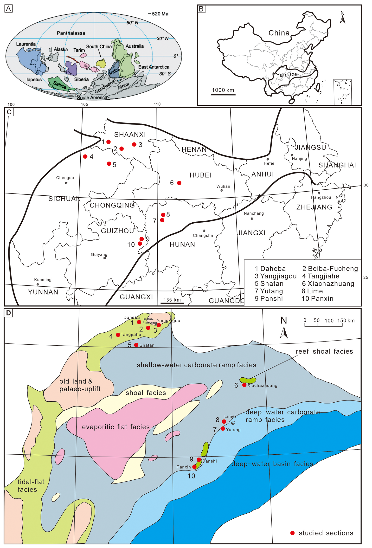
Figure 1Maps showing the sections studied and depositional facies in the Middle–Upper Yangtze Platform region during Cambrian Series 2. (A) Early Cambrian (520 Ma) paleocontinental reconstruction (Li et al., 2021, fig. 1). (B) Location of the Yangtze region within China. (C) Locality map showing the sections studied. (D) Lithofacies paleogeographic map of the Middle–Upper Yangtze region during Cambrian Series 2. Red dots indicate study sections discussed in this paper, after Zhang et al. (2016). Paleogeographic facies adapted from Mou et al. (2012).
The Xiannüdong Formation mainly consists of massive, relatively pure or clastic-rich, oolitic, microbial, and reef limestones, with occasional layers of sandstone and siltstone (Zeng, 2020) (Fig. 2), The Xiannüdong Formation contains a relatively higher amount of terrigenous input. The overlying Yanwangbian Formation has thin- to medium-bedded calcareous siltstone and fine sandstone at its base, overlain by light- to dark-red claystone, siltstone, and fine sandstone. The underlying Guojiaba Formation comprises grayish-green siltstone and claystone with abundant trace fossils. The Xiannüdong Formation (Cambrian Stage 3) is predominantly carbonate with some siliciclastics (Shen, 2015). In the Beiba-Fucheng section of southern Shaanxi, a large reef complex (Fig. 3A) contains Archaeopharetra chengkouensis and calcimicrobes (Epiphyton, Renalcis) as primary reef-building organisms, accompanied by trilobite and echinoderm bioclasts. Its cement contains a minor amount of quartz sand. The middle part of the Shatan section in northern Sichuan contains a prominent reef (Fig. 3B) with well-preserved archaeocyaths exposed on weathering surfaces, together with calcimicrobes and echinoderm and trilobite bioclasts. In the Tangjiahe section, a small reef (Fig. 3C) shows densely packed archaeocyaths on weathered surfaces (Fig. 3D), together with echinoderm and trilobite bioclasts. The Yangjiagou section is predominantly microbial carbonate (thrombolite and stromatolite); a small number of archaeocyaths are visible in the outcrop. In the Daheba Formation, oolite beds are frequently interbedded with fragmented archaeocyaths.
The Tianheban Formation has conformable contacts with the underlying yellowish-green silty shale of the Shipai Formation and with overlying massive dolostones of the Shilongdong Formation (Fig. 2). It is composed of thin- to medium-bedded argillaceous-banded limestone, intercalated with oolitic and conglomeratic limestones and silty shale. The middle–upper intervals of the formation contain archaeocyath-bearing reefs, interpreted as open-platform deposits (Wang, 1986). Microbial-archaeocyath reef facies, well developed in the Xiachazhuang section (Fig. 3E), contain abundant fossils (Fig. 3F) such as Archaeocyathus yichangensis, calcified cyanobacteria, calcimicrobes of uncertain affinity, and bioclasts (trilobites, chancelloriids, echinoderms).
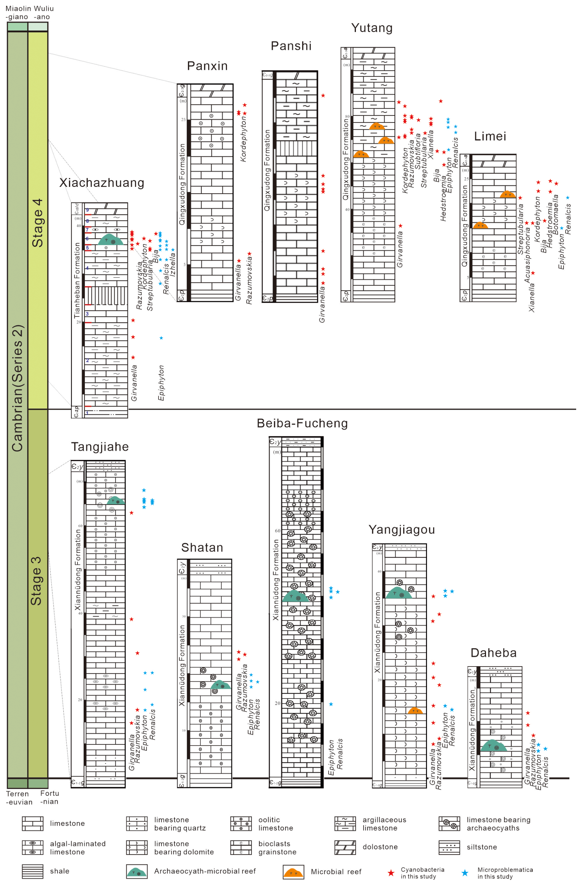
Figure 2Stratigraphic column of 10 shallow marine carbonate sections, both reefal and non-reefal, from Cambrian Series 2 of the Yangtze Platform, showing horizons with calcified cyanobacteria and associated microfossils.
The Qingxudong Formation, conformably overlying the Palang Formation, is composed of alternating layers of gray-green to yellow-green clay-rich shale, light-gray calcareous shale, and silty shale, with thin argillaceous limestone interbeds, transitioning upward into the Gaotai or Aoxi formations (Fig. 2), which consist predominantly of laminated dolostone. The Qingxudong Formation in the Yutang section comprises four members: (i) basal light-gray micrite grading upward into (ii) dark-gray limestone, (iii) light-gray mottled limestone, and (iv) dark-gray to black argillaceous-banded limestone. Within the lower part of the upper argillaceous-banded limestone unit, thick-bedded intervals contain calcimicrobe reef limestone (Fig. 3H). The upper part transitions to thin-bedded argillaceous-banded limestone. In the Panxin section, the lower member of the Qingxudong Formation consists of limestone grading upward into limestone-bearing dolomite, suggesting a tidal-flat environment or an exposure event (inner-ramp facies). The upper member includes limestone intervals of oolitic limestone containing calcified cyanobacteria, indicating transformation into a high-energy bank environment (mid-ramp facies). In the Panshi section, the lower member of the Qingxudong Formation is limestone; the middle member is limestone-bearing dolomite, with its top part missing; and the upper member is argillaceous limestone, reflecting a distal open-platform environment (outer-ramp facies). In the Limei section, the Qingxudong Formation is predominantly limestone. An interval of bioclastic grainstone is present at the base, and limestone-bearing dolomite forms the upper part. Numerous calcified cyanobacteria occur in the upper–middle section.
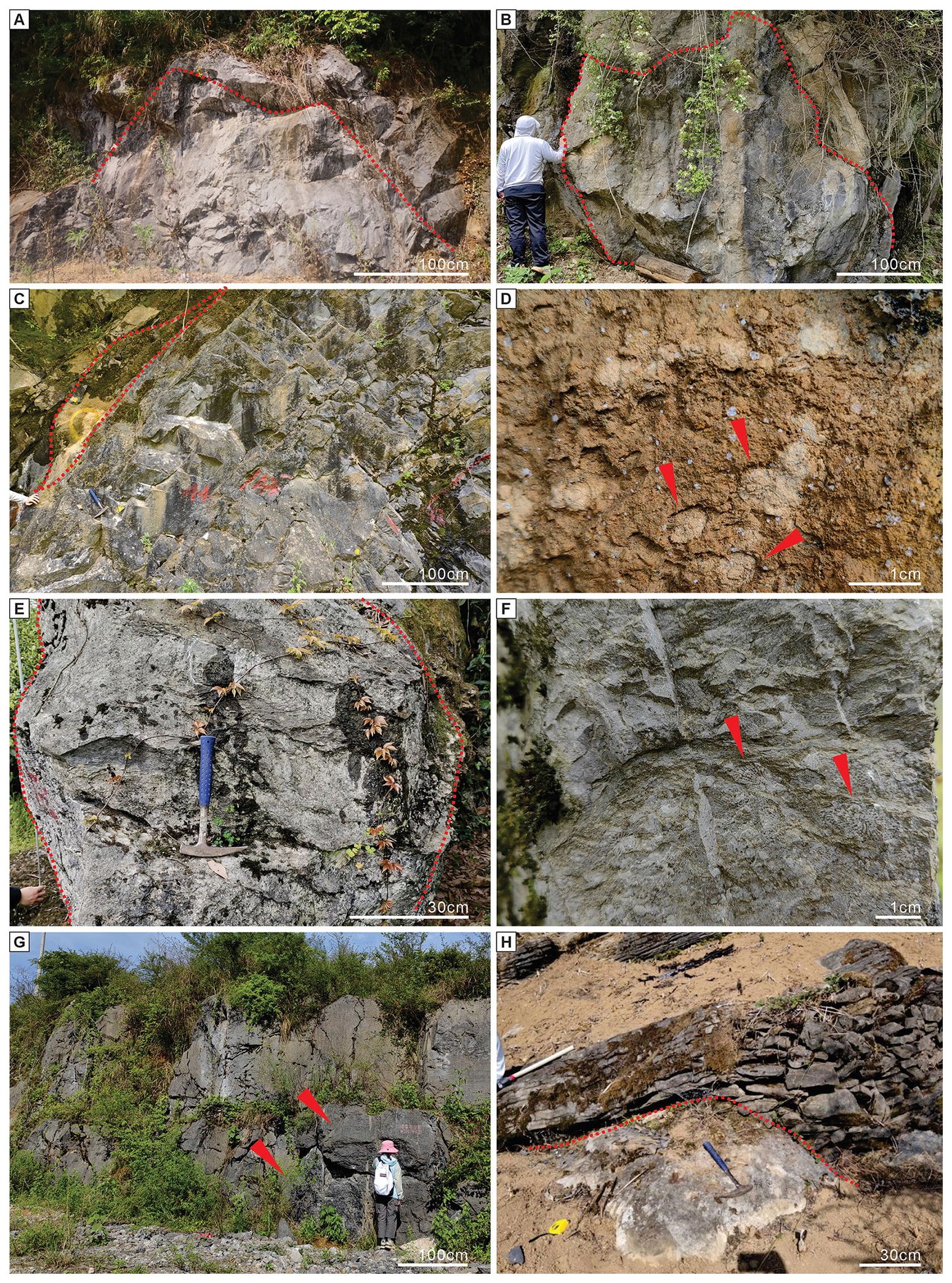
Figure 3Cambrian Series 2 reef outcrops in the sections studied. Large reef (outlined in red) in the Xiannüdong Formation, Fucheng section. (B) Reef (outlined in red) in the lower–middle Xiannüdong Formation, Shatan section. (C) Archaeocyath-bearing reef in the Xiannüdong Formation, Tangjiahe section (small archaeocyath reef outlined in red). (D) Weathered surface of the reef in (C) showing distinct archaeocyaths (red arrows). (E) Calcimicrobe–archaeocyath reef (outlined in red) in the Tianheban Formation, Xiachazhuang section. (F) Weathered surface of the reef in (E) showing archaeocyaths (red arrows). (G) Argillaceous-banded limestone (red arrows), Qingxudong Formation, Panshi section. (H) Dome-shaped calcimicrobial reef (partially exposed) in dark-gray argillaceous-banded limestone (Member 4, Qingxudong Formation), Yutang section.
A total of 13 094 thin sections were prepared from samples collected at 10 outcrop sections and examined using a Canon DSLR camera coupled with an OLYMPUS BX53 transmitted-light polarizing microscope. Detailed microscopic observations of calcified cyanobacteria and problematic calcimicrobial fossils were conducted, with comprehensive documentation and acquisition of over 4000 photomicrographs. Fossil identification was based on diagnostic characteristics, including filament diameter, length and branching, sheath thickness, and overall morphology, following taxonomic criteria based on previous studies (e.g., Debrenne et al., 1991; Riding, 1991a; Liu et al., 2016a, 2021). Systematic classification incorporated both fossil and present-day cyanobacterial features. All specimens and thin sections are housed in the Department of Geology, Northwest University.
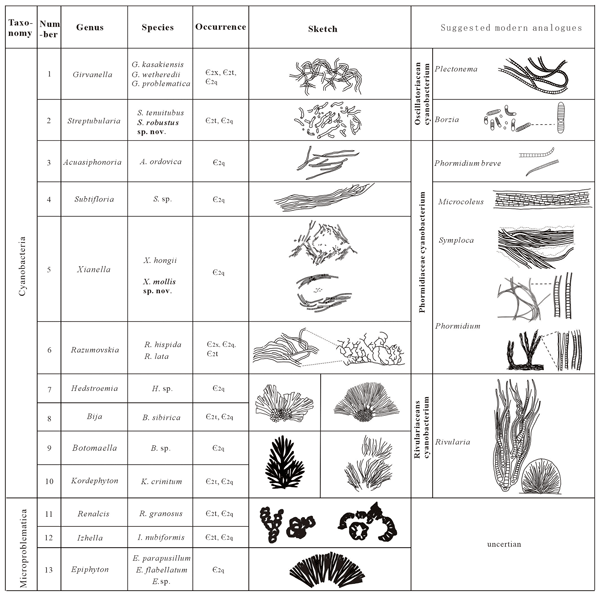
Overall, 20 species in 13 genera of calcified cyanobacteria and associated microfossils, including 2 new species, were identified. Girvanella, Subtifloria, Xianella, Acuasiphonoria, Razumovskia, and Streptubularia are attributed to the Oscillatoriales (cyanobacteria); Hedstroemia, Kordephyton, Botomaella, and Bija are attributed to Nostocales (cyanobacteria). In addition, associated Epiphyton, Renalcis, and Izhella are reported and attributed to calcified Microproblematica. Most of these fossils are from the Qingxudong and Tianheban formations. The locations, horizons, and sedimentary facies of these calcified cyanobacteria and Microproblematica are illustrated in Fig. 2; the taxa are illustrated in Fig. 4.
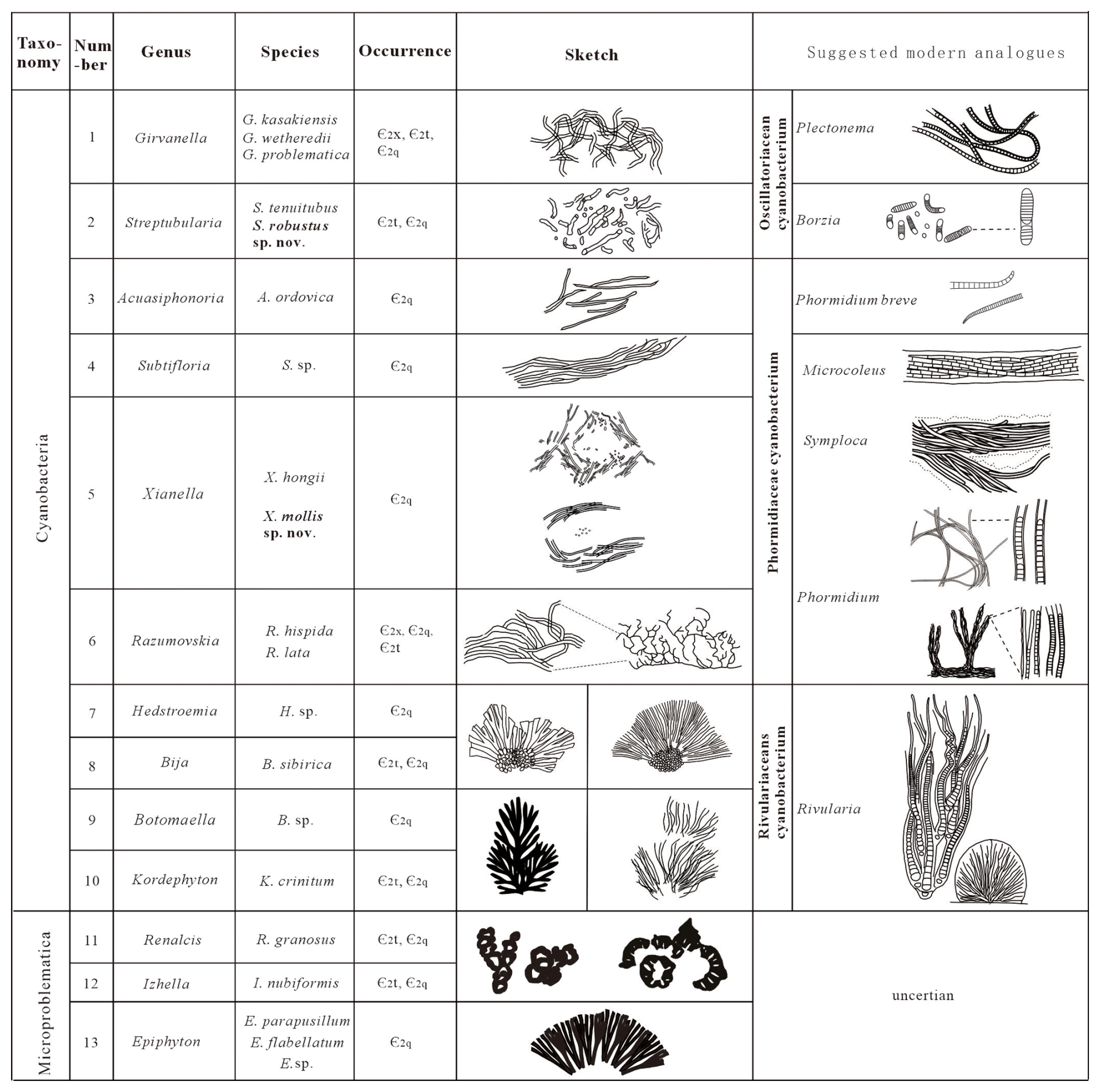
Figure 4Taxa of calcified cyanobacteria and associated calcified Microproblematica recognized in this study, with suggested present-day analogues. Species in bold are new (sketches of fossils and modern analogues are based on Liu et al., 2016a).
-
Cyanobacteria Stanier, 1974
-
Order Oscillatoriales Elenkin, 1949
-
Genus Girvanella Nicholson and Etheridge, 1878
(Fig. 5)
-
1973 Nicholsonia Korde: 212, pl. 43, fig. 3; pl. 44, fig. 1; pl. 45, fig. 1.
Type species. Girvanella problematica Nicholson and Etheridge, 1878; Upper Ordovician, Scotland.
Diagnosis. Calcareous tubular filaments; long and curved; loosely to tightly tangled; micritic thin wall; filament diameters usually range from a few microns to tens of microns.
Comparison. Girvanella is one of the most widely known calcified cyanobacteria of the Paleozoic (Danielli, 1981). Girvanella species are mainly distinguished based on the overall diameter of the filament and the wall thickness. Korde (1973) distinguished Nicholsonia from Girvanella, but Nicholsonia was subsequently considered to be a synonym of Girvanella (Danielli, 1981) due to the lack of difference between some species of Nicholsonia and Girvanella. Batenevia and Botominella (which has been considered to be a synonym of Subtifloria; see Luchinina, 1975) are distinguished from Girvanella by their sarciniform (bundled) filament arrangement.
Affinity. Girvanella is generally regarded to be cyanobacterial (Bornemann, 1886; Pollock, 1918; Frémy and Dangéard, 1935). Riding's (1977b) comparison of it with calcified cyanobacterial sheaths, as in present-day Plectonema, has been widely accepted (Feng et al., 2010; Min et al., 2019, 2024; Liu et al., 2016a, 2021; Zheng et al., 2024; Zhang et al., 2024; Xiao et al., 2025).
Species. Conventionally, species of Girvanella are primarily distinguished by filament diameter and wall thickness (Wood, 1957). Zhang et al.'s (2024) revision of Girvanella species, by combining statistical analysis of their diameters with the taxonomic criteria of Mamet and Roux (1975), recognized four Girvanella species: kasakiensis, problematica, wetheredii, and staminea.
-
Girvanella kasakiensis Maslov, 1949 emend. Mamet and Roux, 1975
(Fig. 5E)
-
1949 Girvanella ducii var. kasakiensis Maslov: 6.
-
1965 Girvanella aff. ducii Chuvashov: 74, pl. 17, fig. 3.
-
1967 Girvanella ducii Wethered; Wray: 34, pl. 7, fig. 5.
-
1975 Girvanella kasakiensis Maslov; Mamet and Roux: 142, pl. 4, fig. 10; pl. 5, figs. 1, 2, 9, 10.
-
2011 Girvanella kasakiensis Maslov; Liu et al.: 495, pl. 1, fig. 3.
-
2016a Girvanella kasakiensis Maslov; Liu et al.: 188, fig. 4A.
-
2021 Girvanella kasakiensis Maslov; Liu et al.: 7, figs. 4.2, 4.3.
Material. Rare in the Xiannüdong Formation (ϵ2x) at the Yangjiagou section, Hanzhong City, southern Shaanxi Province.
Description. Calcareous unbranched tubular filaments, long and loosely tangled; wall thin, micritic; external diameter 23–28 µm, wall thickness 4–6 µm (Fig. 5E).
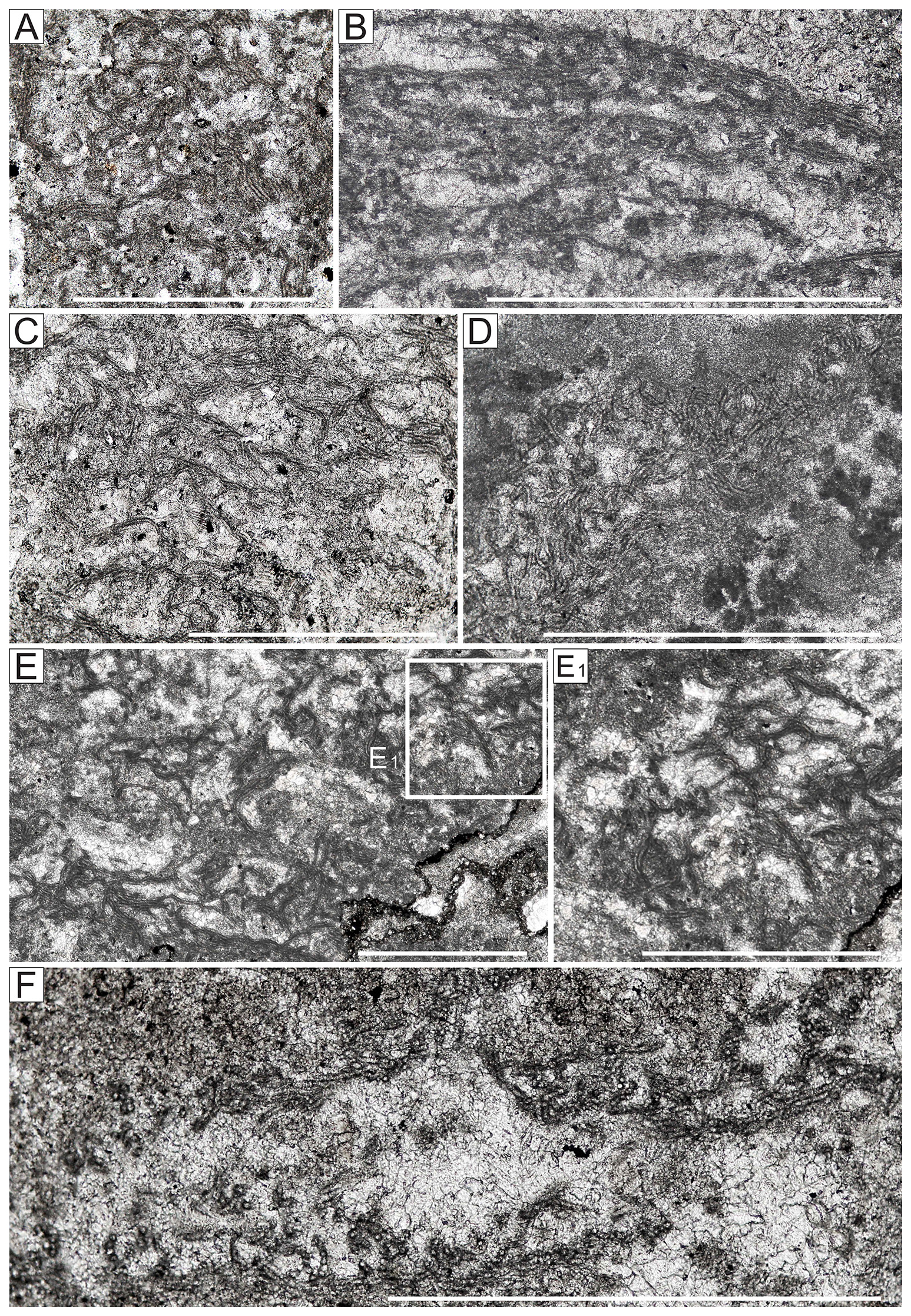
Figure 5Photomicrographs of Girvanella in the early Cambrian of South China. (A–D) Girvanella problematica. (A) Yangjiagou section, sample no. 2-29, oblique longitudinal sections. (B) Xiachazhuang section, sample no. 6-S32, oblique longitudinal sections. (C) Yangjiagou section, sample no. 2-22, oblique longitudinal sections. (D) Xiachazhuang section, sample no. 3-2, oblique longitudinal sections. (E) Girvanella kasakiensis, Yangjiagou section, sample no. 2-6, longitudinal sections. (F) Girvanella wetheredii, Yutang section, sample no. 4-2, longitudinal and cross-sections. All scale bars equate to 1 mm.
-
Girvanella problematica Nicholson and Etheridge, 1878 emend. Wood, 1957(= Girvanella ducii Wethered, 1890
(Fig. 5A–D)
-
1878 Girvanella problematica Nicholson and Etheridge: 23, pl. 9, fig. 24.
-
1890 Girvanella ducii Wethered: 280, pl. 11, fig. 2a–c.
-
1932 Girvanella problematica Nicholson and Etheridge; Høeg: 64, pl. 1, figs. 4–6.
-
1981 Girvanella problematica Nicholson and Etheridge; Bourque et al.: 95, p1. 1, figs. 2–4.
-
2001 Girvanella problematica Nicholson and Etheridge; Riding and Fan: 789, fig. 3B, C.
-
2011 Girvanella problematica Nicholson and Etheridge; Liu et al.: 495, pl. I, fig. 4.
-
2016a Girvanella problematica Nicholson and Etheridge; Liu et al.: 188, fig. 4B.
-
2021 Girvanella problematica Nicholson and Etheridge; Liu et al.: 7, fig. 4.1.
Material. Abundant in the Xiannüdong Formation (ϵ2x) at the Yangjiagou section, southern Shaanxi Province, and in the Qingxudong Formation (ϵ2q) at the Yutang section, Yangtze Platform, Hunan Province, South China.
Description. Calcareous tubular filaments, curved and wound into a loose mass; wall thin, micritic; external diameter 13–22 µm, wall thickness 2–4 µm (Fig. 5A–D).
-
Girvanella wetheredii Chapman, 1908 (= Girvanella incrustans Wethered, 1890, non Bornemann, 1886)
(Fig. 5F)
-
1975 Girvanella wetheredii Chapman; Mamet and Roux: 141, pl. 1, figs. 9–12, pl. 161, pl. 2, figs. 1–5.
-
1995 Girvanella wetheredii Chapman; Mamet and Shalaby: 233, pl. 1, fig. 3.
-
2011 Girvanella wetheredii Chapman; Liu et al.: 495, pl. 1, fig. 1.
-
2016a Girvanella wetheredii Chapman; Liu et al.: 188, fig. 4C.
Material. Locally present in the Qingxudong Formation (ϵ2q) at the Yutang section, Yangtze Platform, Hunan Province, South China.
Description. Calcareous tubular filaments, curved and wound into a loose mass; wall thin, micritic; external diameter 8–12 µm, wall thickness about 1–2 µm (Fig. 5F).
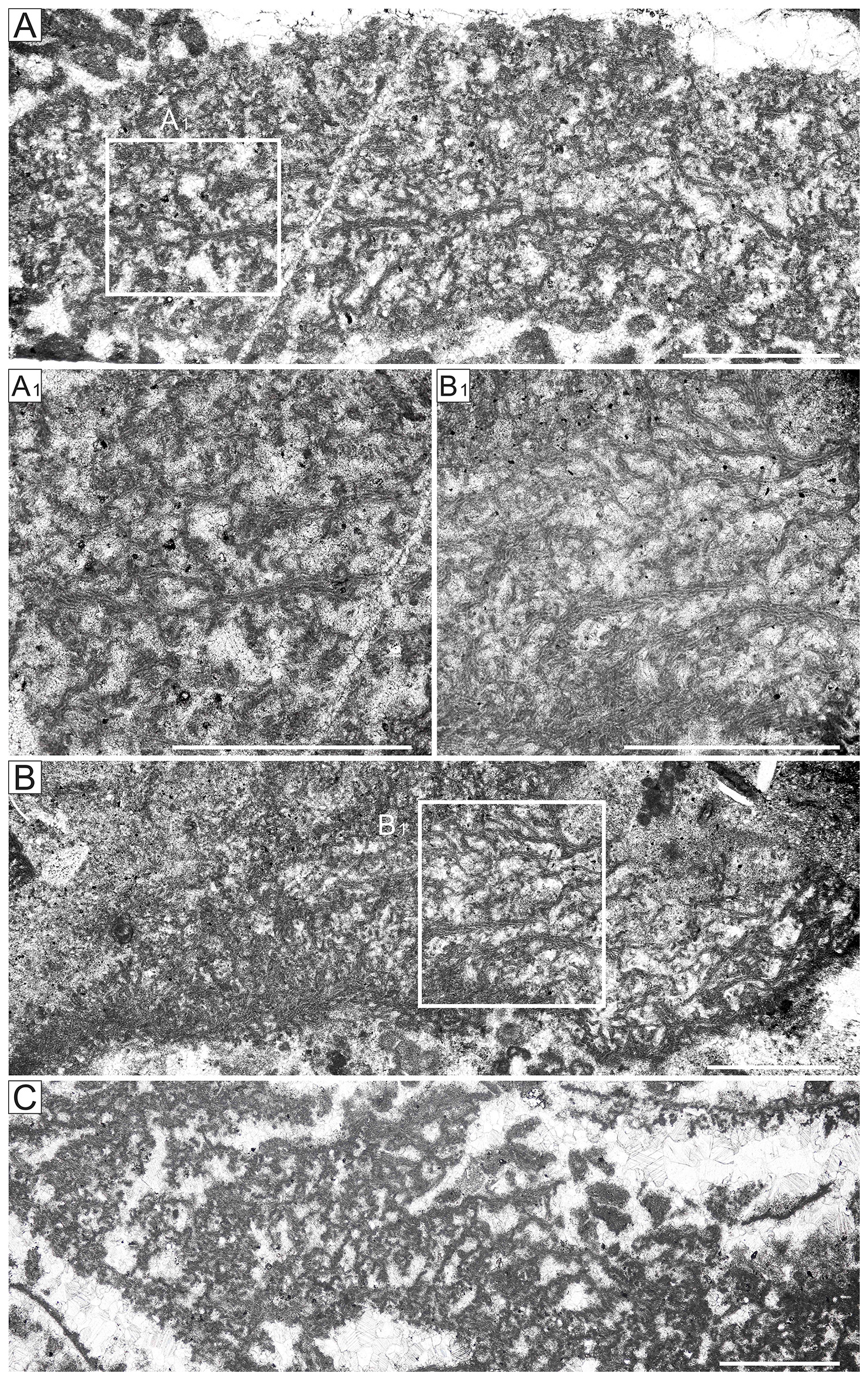
Figure 6Photomicrographs of Razumovskia fossils, early Cambrian, South China. (A–B) Razumovskia hispida. (A) Yangjiagou section, sample no. 2-6. (A1), enlargement of part of (A). (B) Xiachazhuang section, sample no. 6-S04. (B1) Enlargement of part of (B). (C) Razumovskia lata Yangjiagou section, sample no. 2-31. All scale bars equate to 1 mm.
-
Genus Razumovskia Vologdin, 1939
(Fig. 6A–C)
-
1937 Razumovskia Vologdin; Krasnopeeva: 19, pl. III, fig. 24.
-
1990 Trichophyton Bian and Zhou: 6. pl. 4, fig. 8.
-
2016a Razumovskia Vologdin; Liu et al.: 189, pl. 4, fig. G.
Type species. Razumovskia uralica Vologdin in Krasnopeeva, 1937; Lower Cambrian; South Urals, Russia.
Diagnosis. Calcareous tubular filaments, long and curved, arranged in a parallel or vertically alternating pattern, and grouped into a mat; distal tubes can extend vertically from the mat, bending into different lengths; wall thin micritic. Comparison. Razumovskia has been reported from the early Cambrian (Vologdin, 1939; Korde, 1973; Drosdova, 1980; Adachi et al., 2023; Zheng et al., 2024), Late Cambrian (Lee and Riding, 2022) and Late Ordovician (Liu et al., 2016a). Razumovskia and Girvanella are similar in terms of basic filament morphology; the main difference is filament arrangement. Girvanella filaments are commonly irregularly tangled, whereas Razumovskia filaments are typically arranged in parallel or vertically alternating, with a tendency to curve upward at the end. Trichophyton, proposed by Bian and Zhou (1990) and also reported from the Ordovician of the Ordos Basin of North China (Ye et al., 1995), is considered to be a junior synonym of Razumovskia (Liu et al., 2016a).
Affinity. Liu et al. (2016a) compared the filament arrangement in Razumovskia to that of present-day Phormidium.
Species. Korde (1961, 1973) recognized five species based on filament diameter: R. kiyanica, R. multispora, R. seriate, R. hispida, and R. grandis. Razumovskia species recognized in the present study are R. hispida and R. lata, as described by Drosdova (1980).
-
Razumovskia hispida Korde, 1973
-
1973 Razumovskia hispida Korde: 128, pl. 10, fig. 1; pl. 11, fig. 1a.
Material. Widespread in the Xiannüdong Formation (ϵ2x) at the Yangjiagou section, southern Shaanxi Province, and locally present in the Tianheban Formation (ϵ2t) at the Xiachazhuang section, Hubei Province, South China.
Description. Short filaments extending from the mat, diameter 10–15 µm, wall thickness 2 µm (Fig. 6A–B).
-
Razumovskia lata Drosdova, 1980
-
1980 Razumovskia lata; Drosdova: pl. IV, fig. 3; Plate V, figs. 1, 3, 5.
-
1990 Trichophyton changshanensis; Bian and Zhou: 6, pl. 4, fig. 8, IV.
-
1995 Trichophyton sp.; Ye et al: 16, pl. 8, fig. 7.
-
2014 Razumovskia lata; Liu: 57, pl. 4-4, fig. B.
Material. Only observed in the Xiannüdong Formation (ϵ2x) at the Yangjiagou section, Hanzhong City, southern Shaanxi Province.
Description. Short filaments extend from longer and more curved mat-like filaments, with diameter ∼ 8 µm (Fig. 6C).
-
Genus Subtifloria Maslov, 1956
(Fig. 7A)
-
1959 Botominella Reitlinger: 25, pl. 10, figs. 1–7.
-
2009 Girvanella Nicholson and Etheridge; Wang et al.: fig. 2a.
-
2014 Girvanella Nicholson and Etheridge; Rong et al.: fig. 5a, b.
Type species. Subtifloria delicata Maslov, 1956; Lower Cambrian; Siberian Platform, Russia.
Diagnosis. Calcified tubular filaments, nearly parallel and clustered into bundles, uniform filament diameter, thin micritic wall.
Comparison. Calcified Subtifloria has mainly been reported from the early–middle Cambrian (Maslov, 1956; Luchinina, 1975; Luchinina and Terleev, 2003; Mei et al., 2020; Li and Cong, 2025) and the Middle–Late Ordovician (Riding and Fan, 2001; Liu et al., 2016a, 2021; Tang, 2024). Rare examples of Subtifloria have been described from the Mid–Late Devonian in southern China (Feng et al., 2010). Unlike Batenevia, in which the filaments are also in bundles; Subtifloria filaments do not branch. Luchinina (1975) considered Botominella to be a junior synonym of Subtifloria. Subtifloria resembles Girvanella in terms of tube size but differs in terms of the subparallel arrangement of its filaments in bundles and its possible branching. Specimens regarded to be Girvanella from the Middle–Upper Ordovician of the Tarim Basin (Wang et al., 2009; Rong et al., 2014) have since been identified as Subtifloria (Feng et al., 2010; Liu et al., 2016a; Xiao et al., 2025). Subtifloria latissima Luchinina from the Lower Carboniferous of the Kuzbass (Bogush et al., 1990, pl. 3, fig. 1) may be Girvanella (Feng et al., 2010; Liu et al., 2016a; Xiao et al., 2025).
Affinity. Luchinina (in Chuvashov et al., 1987) compared Subtifloria with present-day Microcoleus, which shows filaments arranged in parallel bundles, supporting a cyanobacterial affinity, and this has been endorsed (Feng et al., 2010; Liu et al., 2016a).
Species. Subtifloria currently consists of two species, S. delicata and S. latissima (Bogush et al., 1990). S. delicata filaments can be up to 12 mm long, with an outer diameter of ∼ 27 µm and a wall thickness of around 5 µm; Subtifloria latissima resembles Girvanella; its taxonomic status is uncertain.
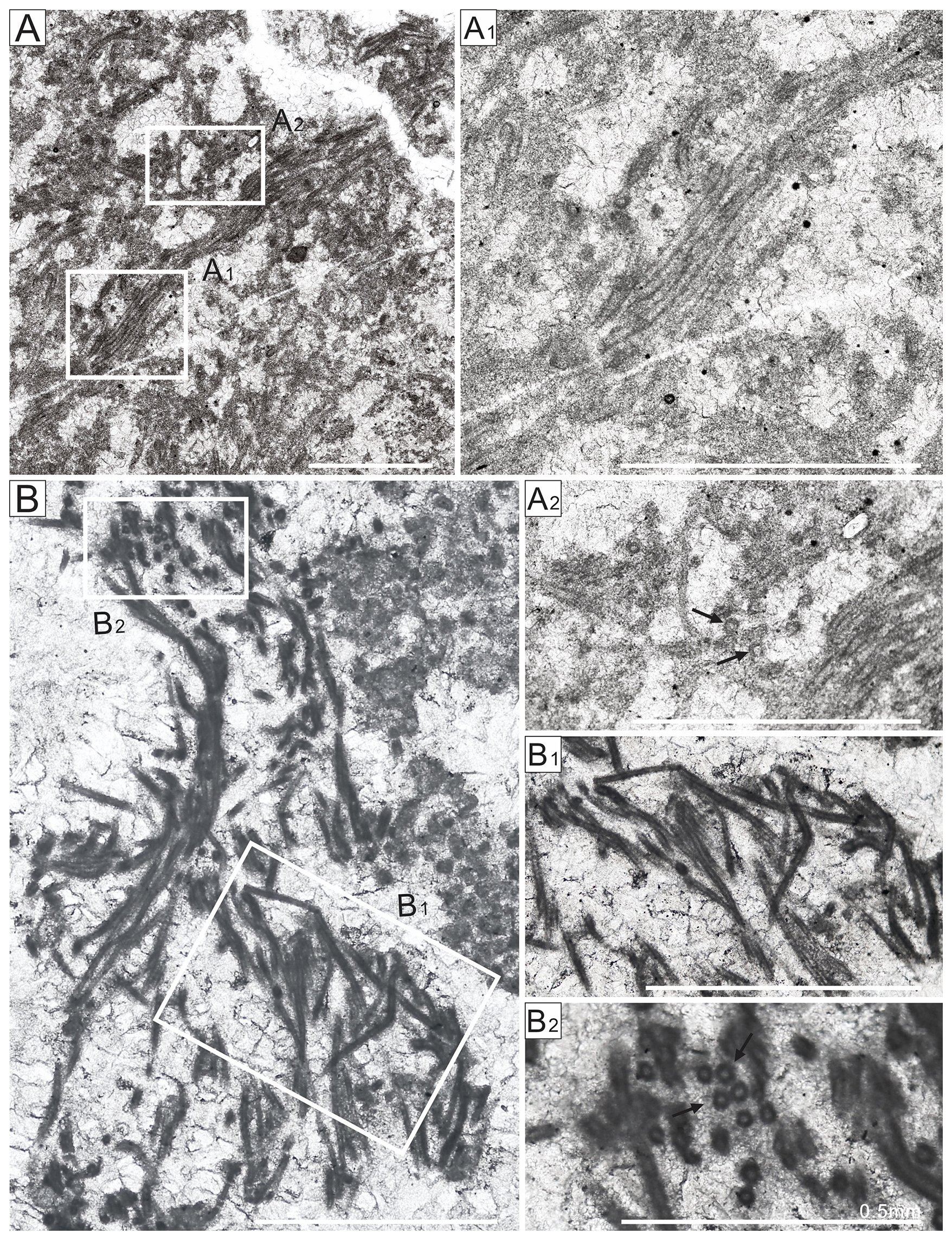
Figure 7Photomicrographs of Subtifloria and Acuasiphonoria fossils, early Cambrian, South China. A, Subtifloria sp., Yutang section, sample no. 5-S17, longitudinal section. (A1) Enlargement of part of (A) longitudinal section. (A2) Enlargement of part of (A). Arrows indicate cross-sections. (B) Acuasiphonoria ordovica, Limei section, sample no. 3-DLC-1, longitudinal section. (B1) Enlargement of part of (B) longitudinal section. (B2) Enlargement of part of (B). Arrows point to cross-sections. All scale bars equate to 1 mm, except where indicated.
-
Subtifloria sp.
Material. Rarely present in the Qingxudong Formation (ϵ2q) at the Yutang section, Hunan Province, South China.
Description. Filaments, almost parallel and clustered into bundles, with external diameter 34–40 µm.
Remarks. Due to its unusually large diameter, much greater than currently recognized Subtifloria species (Fig. 7A), its classification remains uncertain.
-
Genus Acuasiphonoria Liu et al., 2016
(Fig. 7B)
-
1973 Kordephyton Korde: 289, pl. 13, fig. 2; pl. 14, figs. 2, 3.
Type species. Acuasiphonoria ordovica Liu et al., 2016; Upper Ordovician, Katian, Lianglitag Formation; Tarim Basin, Xinjiang Province, Northwest China.
Diagnosis. Straight to gently curved, calcified long tubular filaments, appearing to end in a sharp point; possibly branched at an acute angle, tube wall micritic.
Comparison. Acuasiphonoria has previously been reported from the Late Ordovician (Liu et al., 2016a, 2021); this is its first report from the Cambrian. Acuasiphonoria is distinguished from Girvanella and Subtifloria by long straight or only slightly curved filaments that appear to taper, needlelike. Acuasiphonoria can resemble branched, bushy forms of Proaulopora in its macroscopic morphology. However, Acuasiphonoria is morphologically distinct from Tubophyllum and the Proaulopora Group (Proauloporaceae) as a whole since it lacks a multilayered wall structure and distinctive whorl-like collars which are characteristic of the Proaulopora Group. Moreover, its sizes differ considerably: members of the Proaulopora Group are much larger, generally 50–100 µm in diameter, whereas the filament diameter of Acuasiphonoria is 22–24 µm.
Affinity. Acuasiphonoria is interpreted as an oscillatoriacean sheath (Liu et al., 2016a). By terminating in a sharp point, Acuasiphonoria is similar to Phormidium breve (Phormidiaceae), whose trichomes similarly show abrupt apical attenuation (Fig. 4).
Species. Acuasiphonoria currently consists of only one species, Acuasiphonoria ordovica.
-
Acuasiphonoria ordovica Liu et al., 2016
-
2016a Acuasiphonoria ordovica Liu et al., p. 191, fig. 5A–C.
-
2021 Acuasiphonoria ordovica Liu et al.: 6, pl. 4, figs. 6–7.
Material. Rarely present in the Qingxudong Formation (ϵ2q) of the Limei section, Yangtze Platform, Hunan Province, South China.
Description. Elongate straight, separated cylindrical filaments, slightly curved, ending in a sharp point; extending straight for up to 1 mm; external diameter 22–24 µm; wall thickness 4–6 µm, micritic (Fig. 7B2).
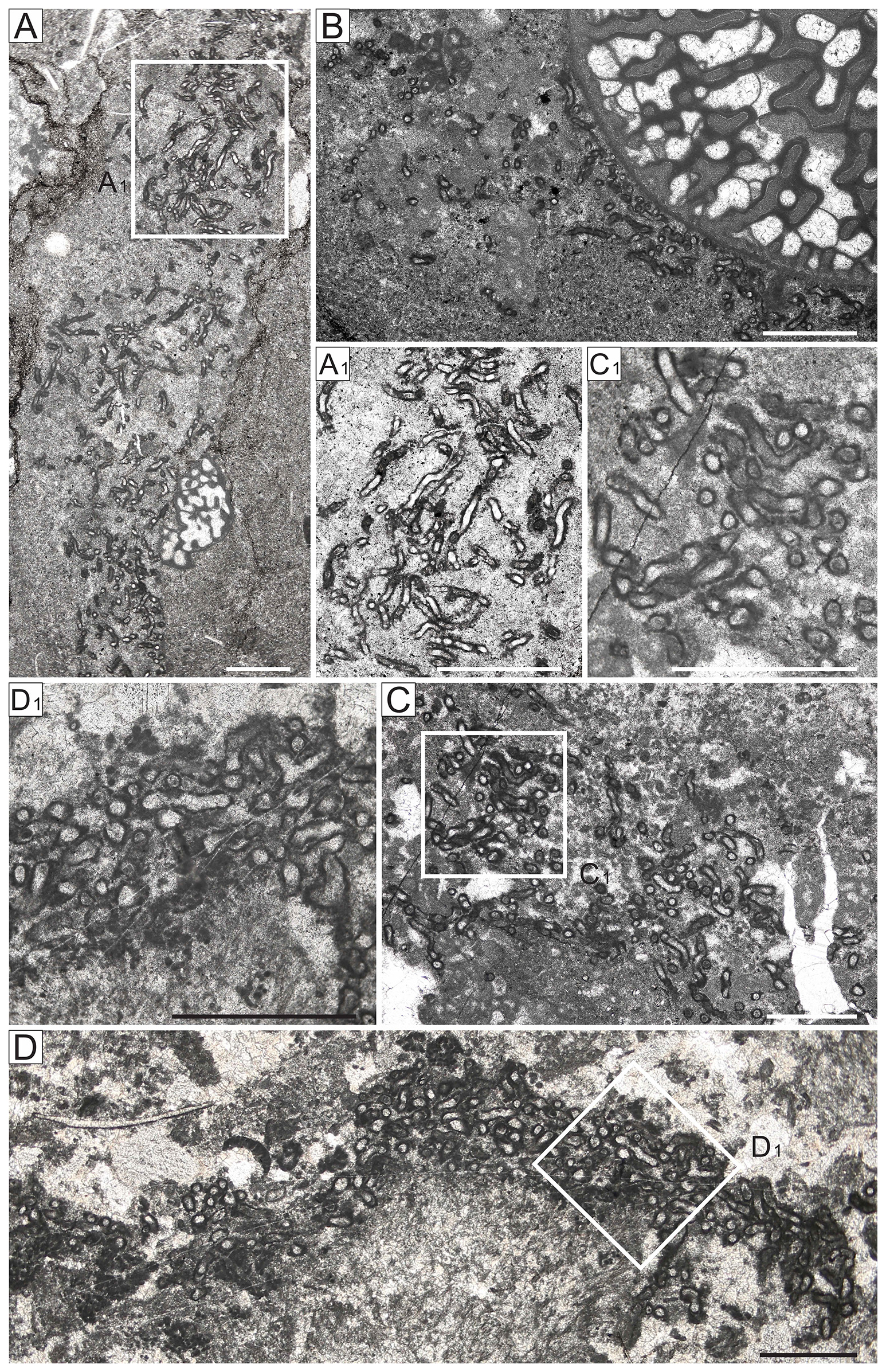
Figure 8Photomicrographs of Streptubularia, early Cambrian, South China. (A–B) Streptubularia tenuitubus; (A) Streptubularia tenuitubus sp. et gen. nov., Xiachazhuang section, sample no. 6-S02 longitudinal section and cross-section. (A1) Enlargement of part of (A). (B) Streptubularia tenuitubus with archaeocyath and Renalcis, Xiachazhuang section, sample no. 6-1. (C–D) Streptubularia robustus n. sp.; (C) Streptubularia robustus n. sp., longitudinal section and cross-section. (D) Streptubularia robustus sp. nov., Yutang section, sample no. 5-S03, longitudinal section and cross-section. D1 Enlargement of part of (D). All scale bars equate to 1 mm.
-
Genus Streptubularia Zheng, Xiao, and Liu, 2025
Type species. Streptubularia tenuitubus Xiao et al., 2025; Cambrian, Series 2, Jianchang and Zhushadong Formation, Liaoning and Shandong provinces, North China.
Diagnosis. Robust curved micritic tube with thick micritic wall; length-to-width ratio 3:1 to 6:1; irregularly constricted; Y-shaped dichotomous branching.
Comparison. Streptubularia has previously been reported from the early Cambrian of North China (Xiao et al., 2025). In morphology, Streptubularia is somewhat similar to Girvanella Nicholson and Etheridge, 1878, but is unusually large. Lee et al. (2014) reported a large-diameter Girvanella-like fossil. However, in addition to being large, Streptubularia shows branching. A species of Nicholsonia, N. grandis, can show branching and reach a diameter of 50 µm (Korde 1973, p. 212), closely resembling Streptubularia. Nicholsonia was considered to be a synonym of Girvanella (Danielli, 1981, p. 96) due to an apparent lack of difference in illustrations (Korde, 1973, pl. XLV). Nonetheless, Nicholsonia does appear to exhibit morphological differences among its constituent species, although its features are difficult to recognize from the original illustrations. We suggest that Nicholsonia may be a composite taxon, possibly a heterogeneous association of Girvanella and other organisms. Further work is required to elucidate the distinction between Streptubularia and Nicholsonia.
Affinity. Xiao et al. (2025) compared Streptubularia from the early Cambrian Jianchang Formation in the North China Plate with extant Borzia (Borziaceae, Oscillatoriales), citing short filaments with cross-wall constrictions (Hu et al., 2006). However, Streptubularia is larger than Borzia and resembles a calcified sheath, not a cellular filament. We suggest that Streptubularia could be an oscillatoriacean sheath, as for Girvanella.
Species. Two species of Streptubularia are distinguished, based on tube diameter, relative thickness of the wall, and growth habit.
-
Streptubularia tenuitubus Zheng, Xiao, and Liu, 2025
(Fig. 8A–B)
-
1973 Nicholsonia grandis Korde: 215, pl. 45, fig. 1.
-
2014a Girvanella Adachi et al.: 709, pl. 6, fig. C.
-
2014b Girvanella Adachi et al.: 51, pl. 6, figs. E, F.
-
2025 Streptubularia tenuitubus Xiao et al.: 13, pl. 6 figs. A–E.
Material. Holotype: thin section NWU XCZS02-2-33 from the Xiachazhuang Section (Tianheban Formation, Cambrian Series 2 Stage 4), Yangtze Platform, Hubei, South China.
Description. Curved short tubes, external diameter 60–75 µm, tube length 300–600 µm, micritic wall, internal spar-filled. Often occurs in association with archaeocyaths and the calcified Microproblematica Renalcis and Epiphyton (Fig. 8A–B).
-
Streptubularia robustus sp. nov.
(LSIDurn:lsid:zoobank.org:act:1D577D03-C9B1-4D60-9DBD-071B5B017F7A)
(Fig. 8C–D)
Etymology. robustus; robust.
Material. Holotype: thin section NWU YTS03-1-15 from Yutang section; Cambrian Series 2 Stage 4, Qingxudong Formation; Yangtze Platform, Hunan Province, South China. Abundant in the Limei and Yutang sections.
Description. Curved calcareous thick tubes, external diameter 85–125 µm, wall thickness 9–18 µm, micritic wall (Fig. 8C–D).
Remarks. Streptubularia often occurs in association with Kordephyton. Adachi et al. (2014a) referred to filaments up to 100 µm in diameter to large Girvanella. Similar filaments were described by Lee et al. (2014) and Adachi et al. (2023) as “tubiform microbe” without addressing their systematic position. These fossils resemble Streptubularia.
-
Genus Xianella Lee and Riding, 2016
(Fig. 9A–C)
Type species. Xianella hongii Lee and Riding, 2016; Upper Ordovician Beiguoshan Formation, Ordos Basin, Northwest China.
Diagnosis. Calcareous microfossil; narrow unbranched tubular filaments forming prostrate and erect anastomosing cable-like strands; wall micritic.
Comparison. Xianella has been reported from the Ordovician (Lee and Riding, 2016; Liu et al., 2021) and the early Cambrian of North China (Xiao et al., 2025). The filaments of Xianella form wide cord-like threads, similarly to Cladogirvanella Ott, 1966, with the difference being that Xianella tubes are larger in diameter and are arranged both horizontally and vertically. Bundled filamentous structure also occurs in Subtifloria Maslov, 1956, and in similar genera such as Botominella Reitlinger, 1959, and Batinevia Korde, 1966, but these are not known to form such long branching cables. Ordovician Acuasiphonoria Liu et al., 2016, also consists of arrays of elongate filaments. However, its long, gently curved tubes are pointed and possibly branch at acute angles (Liu et al., 2016a). These features and the lack of anastomosed bundles distinguish Acuasiphonoria from Xianella, which has narrow, unbranched, thin, single-layered-wall tubules (20–45 µm); forms cable-like strands and cavities; and lacks whorl collars. The Proaulopora Group, in general, exhibits thick filaments (up to 70 µm) with multilayered walls, whorl collars, minor branching, and clustered growth.
Affinity. As with Girvanella, Xianella has been interpreted as the calcified sheath of filamentous cyanobacteria (Lee and Riding, 2016; Liu et al., 2021; Xiao et al., 2025). Based on its cable-like strands composed of filament bundles, Xianella was compared by Lee and Riding (2016) with present-day mat-forming cyanobacteria with multiple trichomes in a common sheath, such as Microcoleus. Our specimens support this interpretation (Fig. 11C-a).
Species. The genus is monospecific.
-
Xianella hongii Lee and Riding, 2016
(Fig. 9A–C)
-
2016 Xianella hongii Lee and Riding, p. 4, figs. 4, 5.
-
2021 Xianella hongii Lee and Riding; Liu et al.: 9, pl. 5, figs. 1–6.
Material. Abundant in the Qingxudong Formation (ϵ2q) of the Yutang section, Hunan Province, South China.
Description. Filaments aggregated into mats, inter-filament cavities of varying sizes, cavity diameters range from 100 to 1000 µm, tube diameters range from 20 to 30 µm (Fig. 9A–C).
-
Xianella mollis sp. nov.
(LSIDurn:lsid:zoobank.org:act:B9A502AA-54DC-4117-B7DE-181101E02DA5)
(Fig. 10A–B)
Etymology. Mollis (Latin) means soft.
Material. Holotype: thin section NWU YT S18-2-13 Qingxudong Formation (Cambrian Series 2 Stage 4) Yutang section; Yangtze Platform, Hunan Province, South China. X. mollis occurs locally in the Yutang section.
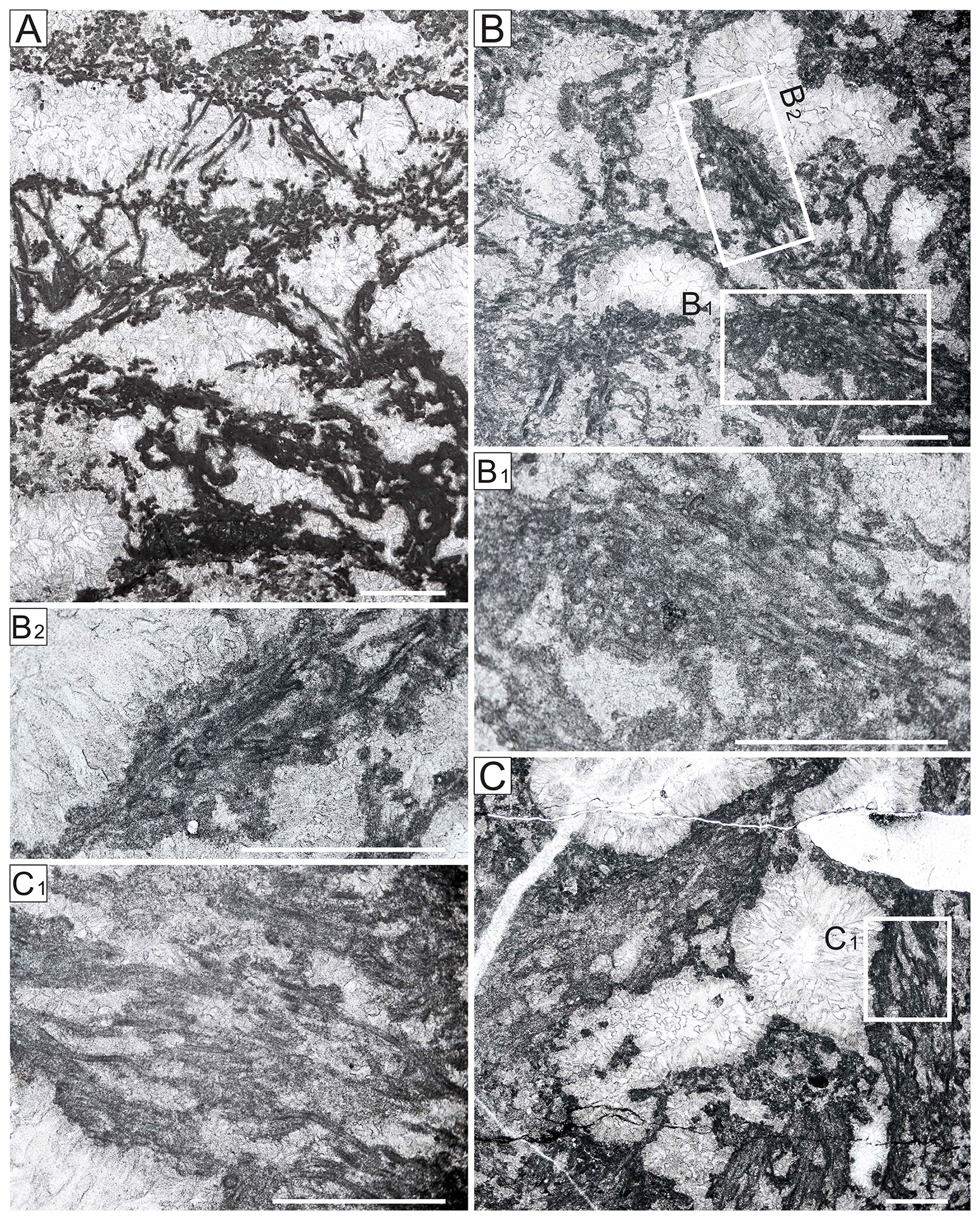
Figure 9Photomicrographs of Xianella hongii, early Cambrian, South China. (A) Overall morphology, Limei section, sample no. 3-DLC-1, longitudinal sections and cross-sections. (B) Medium-sized fenestrae bounded by bundles of Xianella filaments, Yutang section, sample no. 5-S18. (B1) Enlargement of part of (B), cross-section. (B2) Local enlargement of (B), longitudinal section. (C) Yutang section, sample no. 5-S19. (C1) Enlargement of part of (C), longitudinal section. All scale bars equate to 1 mm.
Description. Calcified tubes, unbranched, straight to slightly curved, or wavy. Locally subparallel (Fig. 10A–B). Wall micritic, thick. Wall thickness of 5–10 µm, tubes usually 200–600 µm in length, external diameter 25–45 µm, forming filament bundles with widths of 500–800 µm and lengths up to 3.3 mm; filament bundles can be curved into a U shape (Fig. 10A–B). Mat-like growth.
Remarks. Filaments of X. mollis and X. hongii are very similar. The main distinction is that, in X. hongii, the bundles of filaments usually enclose spar-filled cavities (Lee and Riding, 2016), whereas, in X. mollis, they typically show mat-like growth, and the bundles of filaments are characteristically curved into a U shape and lack anastomose strands (Fig. 11A, C).
The characteristic subparallel arrangement of the tubes in filament bundles somewhat resembles that of Subtifloria (Fig. 11B). Xianella mollis is distinguished by its thick mat-like filament arrangement and concentric annular cross-section (Fig. 11A). X. mollis tubes are similar in size to those of Girvanella and Pachytibia Zheng, Xiao, and Liu, 2025, from the early Cambrian of North China but are distinguished from Girvanella by being more straight. Pachytibia has thicker walls and lacks a subparallel filamentous structure (Fig. 11D) (Xiao et al., 2025).
Affinity. The dense and subparallel arrangement of mat-like filaments in X. mollis can be compared with present-day Phormidiaceae Kützing ex Gomont 1893, such as Symploca and Phormidium. The filament bundles of Symploca can form intertwined, entangled, or parallel aggregates of filaments like those of X. hongii (Fig. 11C-b), whereas, in Phormidium, the bundles of filaments typically have a curved growth pattern similar to that of X. mollis (Fig. 11A). We interpret X. mollis to be the calcified sheath of a cyanobacterium and tentatively assign it to the Phormidiaceae.
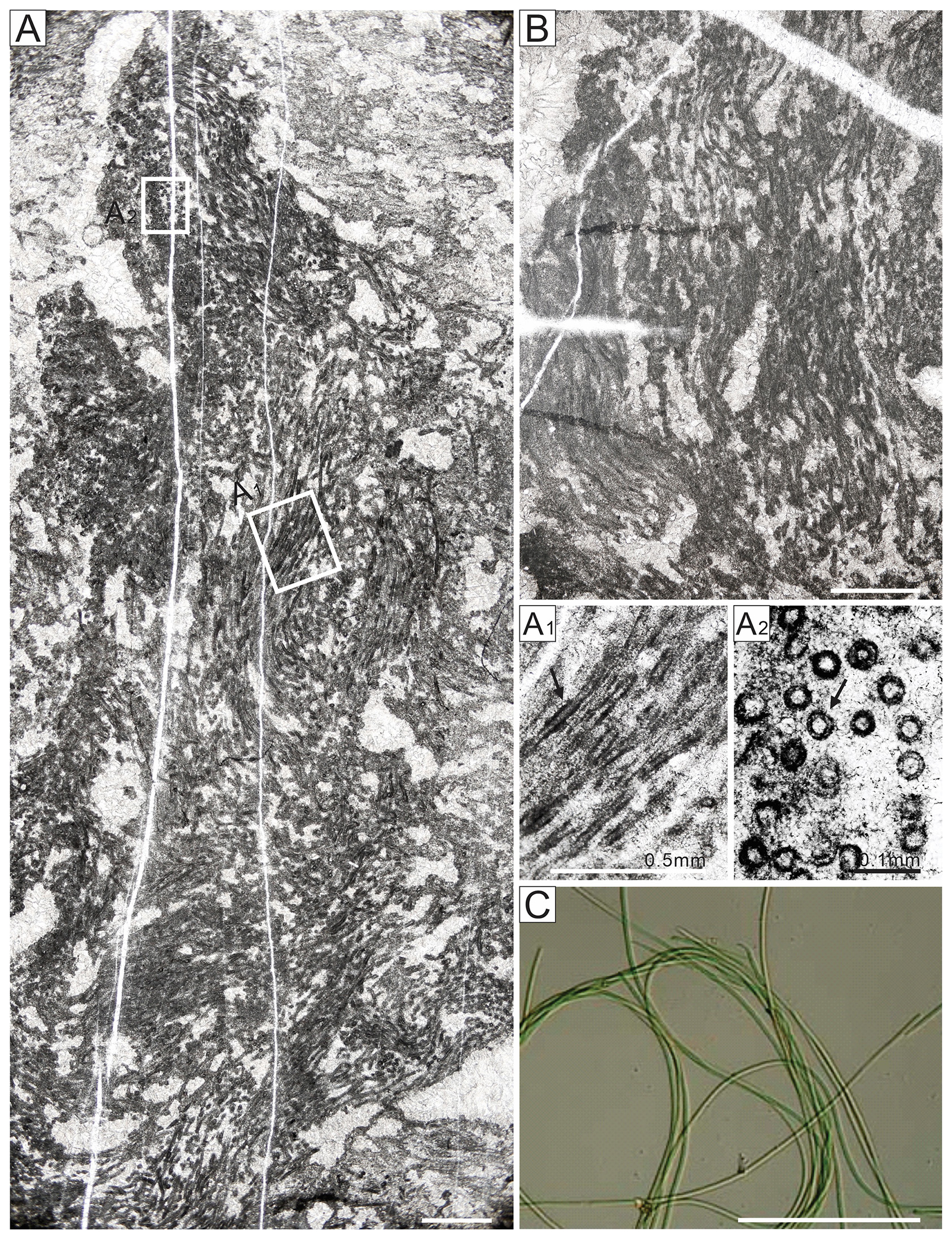
Figure 10Photomicrographs of Xianella mollis sp. nov., early Cambrian, South China. (A–B) Xianella mollis sp. nov. A, overall morphology, Yutang section, sample no. 5-S18, longitudinal sections and cross-sections. (A1) Enlargement of (A), micritic wall (arrowed), longitudinal sections. (A2) Enlargement of (A), concentric ring-like structure (arrowed), cross-sections. (B) Yutang section, sample no. 5-S19, longitudinal section. (C) Extant Phormidium sp. Scale bars equate to 1 mm, except where indicated.
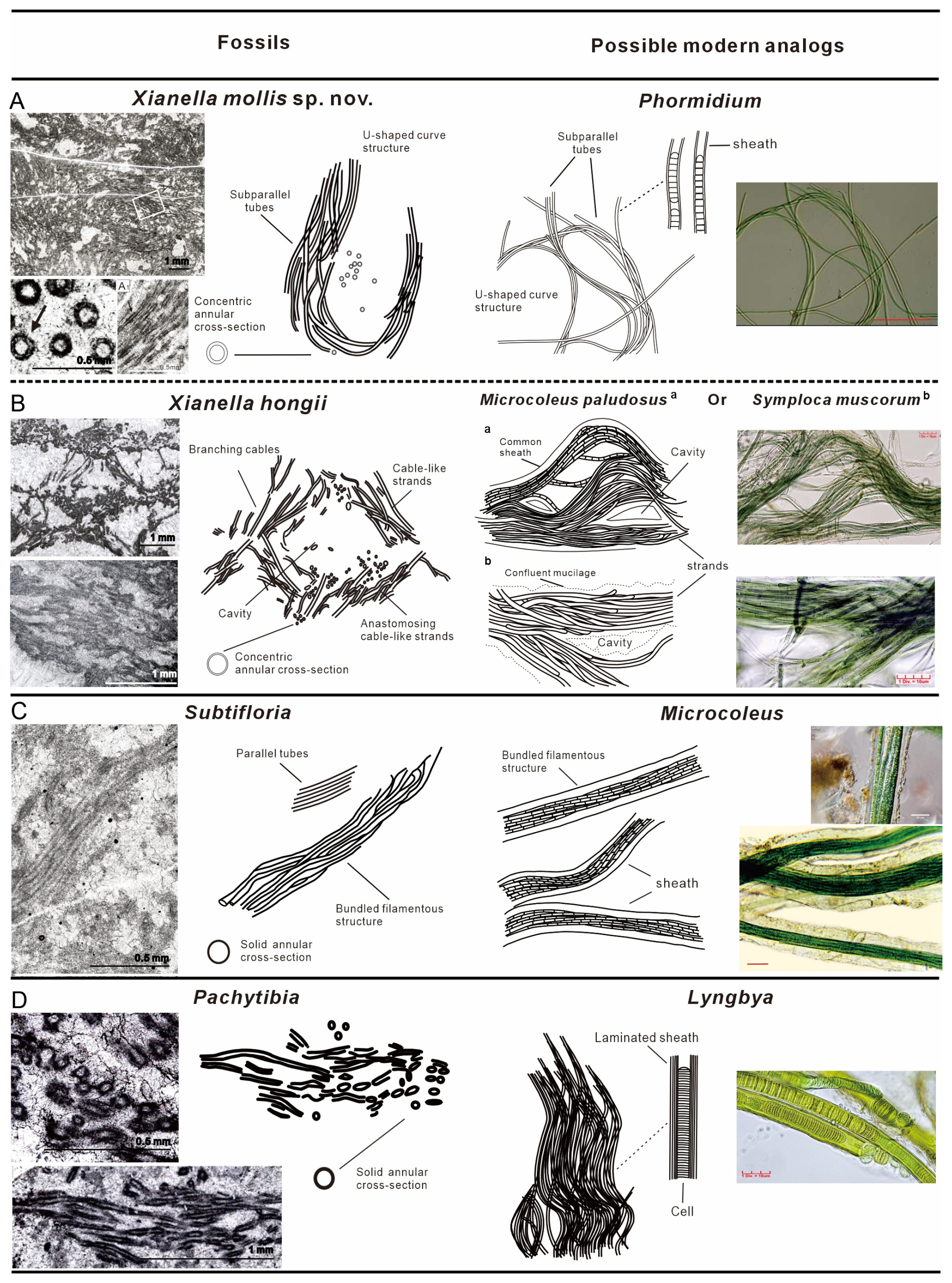
Figure 11(A–D) Xianella mollis sp. nov. compared with taxa such as Xianella hongii, Subtifloria, and Pachytibia from the early Cambrian and possible extant morphological analogs, namely Phormidium, Microcoleus, Symploca (Phormidiaceae, Cyanobacteria), and Lyngbya (Oscillatoriacean, Cyanobacteria). Photomicrographs and sketches: (A) Xianella mollis sp. nov. from this study and present-day Phormidium sp.; arrows indicate U-shaped structure in the fossil and Phormidium. (B) Xianella hongii, extant Microcoleus paludosus, and Symploca muscorum; arrows indicate cable-like strands, branching cables, and cavities. (C) Subtifloria and extant Microcoleus. (D) Pachytibia (from Xiao et al., 2025) and extant Lyngbya. Note the laminated sheath in Lyngbya. Photographs of present-day species are from https://www.atlasofcyanobacteria.com (last access: 11 June 2026)
-
Order Nostocales (Borzi) Geitler, 1925
-
Genus Kordephyton Radugin and Stepanova, 1964
(Fig. 12)
-
1973 Kordephyton Korde: 289, pl. 13, fig. 2; pl. 14, figs. 2, 3.
Type species. Kordephyton crinitum Korde, 1973; Middle Cambrian (Amgan), Elanskoe Formation, Elanskoe, Lena River, Sakha (Yakutia), Russia.
Diagnosis. Thin micritic filaments, long and delicate, in a fan-like pattern. Most filaments appear to be solid; tubular structure is observed in a few micritic filaments and shows relatively thick micritic walls (Fig. 12B).
Comparison. Kordephyton is currently only confirmed from the Cambrian (Riding and Voronova, 1984; Latham and Riding, 1990; Mankiewicz, 1992; Elicki, 1999; Adachi et al., 2023, 2014a, b), although possible occurrences have been reported from the Ordovician (Li et al., 2015). Kordephyton shares a general overall appearance with rivulariacean-like calcified microfossils (e.g., Zonotrichites, Ortonella, Botomaella), characterized by a delicate, micritic, tubular, branched filament arrangement in fan-like radiating clusters, mutually distinguished by details of their filaments, branching patterns, thallus shape, and size. For example, Kordephyton differs from Botomaella in having more slender tubular filaments, with relatively thick micritic walls and a large overall thallus size. Kordephyton's layered, bush-like structure resembles that of some Epiphyton species, and Riding and Voronova (1985) regarded both genera to share “dendroid” morphology. However, the delicate filaments of Kordephyton differ from those of Epiphyton, in which the filaments are generally both wider and shorter.
Affinity. Riding (2001) placed Kordephyton, together with Bija, Bajanophyton, and Botomaella, in the Botomaella Group based on morphological characteristics and supported their likely cyanobacterial affinity. We suggest that Kordephyton is comparable in terms of organization and appearance with some rivulariaceans based on delicate filaments with relatively wide sheaths.
Species. Criteria for distinguishing Kordephyton species include differences in filament diameter, morphology, branching, and shape of notional protosporangia. Three species of Kordephyton, described or revised by Korde (1973), are the most widely recognized (Mankiewicz, 1992): K. crinitum (type species), K. crispum (Korde, 1961) from Kuznetsk Alatau, and K. conglutinatum (Korde, 1973) from East Sayan. In addition, we recognize Kordephyton australis, characterized by continuous, non-laminated filaments, from the Middle Cambrian (Ordian) of the Douglas River, Northern Territory, Australia (Kruse and Reitner, 2014), as a distinct species.
-
Kordephyton crinitum Korde, 1973
(Fig. 12A–D)
Material. Abundant in the Qingxudong Formation (ϵ2q) at the Yutang section, Hunan Province, and locally present in the Tianheban Formation (ϵ2t) of the Xiachazhuang and Qingxudong formations (ϵ2q) at the Limei sections, Yangtze Platform, South China.
Description. Long thin filaments, densely arranged in fan shapes and with blurred filament outlines, can be difficult to mutually distinguish (Fig. 12A–D); branches occur distally on filaments; basal protuberances are tentatively interpreted as putative holdfast structures; protosporangial structures are absent.
Remarks. Discontinuities between constituent filaments and layered bushes, in this species as in other Siberian species, distinguish it from Kordephyton australis (Kruse and Reitner, 2014). Due to often blurred outlines, it can be difficult to confirm that filaments branch and also to distinguish inferred protosporangial structures. The fine and delicate filaments in our specimens are distinct from the coarser filaments typical of K. conglutinatum and resemble K. crinitum (Korde, 1973).
-
Genus Bija Vologdin, 1932
Type species. Bija sibirica Vologdin, 1932; Lower Cambrian; Siberian Platform, Russia.
Diagnosis. Basal cross-section rounded to polygonal, composed of closely spaced long tubular filaments, filaments radially dispersed from the base; irregularly dichotomously branched from one another, with a tendency for filament diameter to decrease distally.
Comparison. Bija is known from the Cambrian (Vologdin, 1932) and Late Ordovician (Liu et al., 2016a). It shows similarities with Hedstroemia Rothpletz, 1913, both having filaments that are circular–polygonal in cross-section. Riding (1991b) suggested that Bija may be a junior synonym of Hedstroemia, differing only in that Bija filaments are longer, and their diameters are less uniform.
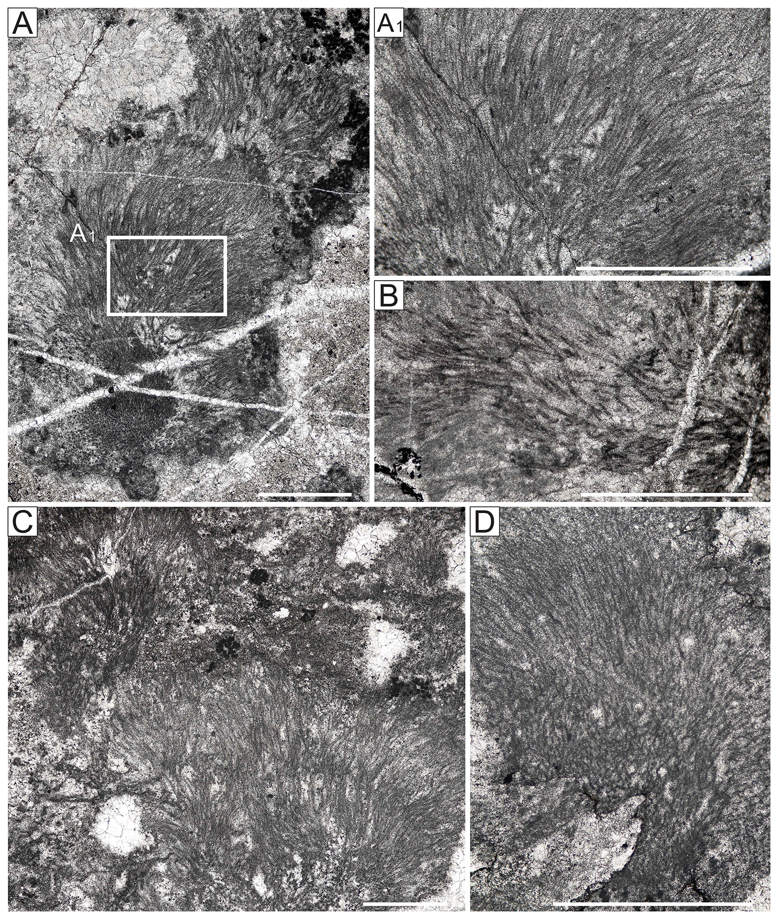
Figure 12Photomicrographs of Kordephyton, early Cambrian, South China. (A–D) Overall morphology, Yutang section, sample no. 5-S08, longitudinal sections and cross-sections. (A1) Enlargement of part of A, radial filaments in longitudinal section. (B) Possible tube structure, Yutang section, sample no. 5-S03, longitudinal section. (C) Radial filaments with Epiphyton (darker micritic masses), Yutang section, sample no. 5-7, longitudinal section. (D) Radial filaments, Yutang section, sample no. 5-7, longitudinal section. All scale bars equate to 1 mm.
Affinity. Bija was placed in the Cyanobacteria by Luchinina (1975). Potential synonymy with Hedstroemia implies shared affinity and comparison with Rivulariaceae (Riding, 1991b). Nonetheless, facies analysis (Liu et al., 2016b, 2017) suggests ecological disparity: Bija is reef- and/or bank-adapted, whereas Hedstroemia is lagoon-adapted. Such environmental differences could suggest distinct ecomorphs or taphonomic effects.
Species. Based on filament diameter and angle of branching, Bija is divided into two species: Bija grandis (Korde, 1973) and Bija sibirica (Vologdin, 1932).
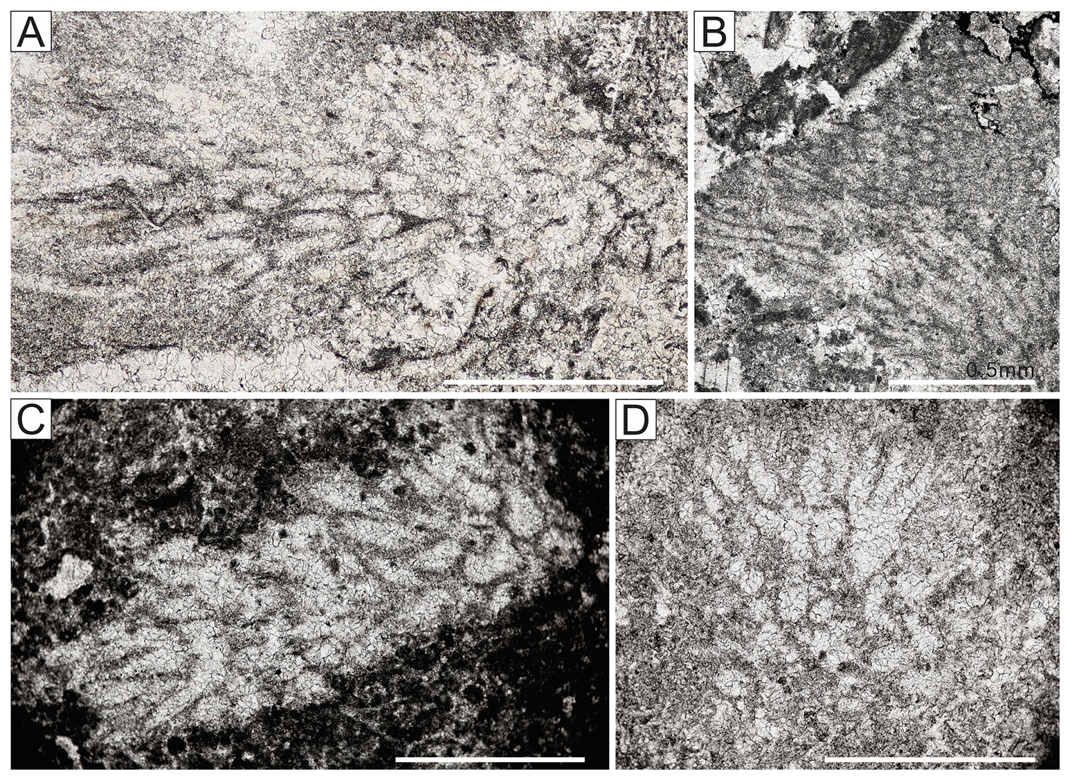
Figure 13Photomicrographs of Bija and Hedstroemia, early Cambrian, South China. (A–B) Bija sibirica. (A) Overall morphology, Yutang section, sample no. 4-4, longitudinal sections and cross-sections. (B) Yutang section, sample no. 5-5, longitudinal sections and cross-sections. (C) Possible Hedstroemia, Yutang section, sample no. 3-11, oblique longitudinal sections. (D) Poorly preserved Hedstroemia, Yutang section, sample no. 4-2. All scale bars equate to 1 mm.
-
Bija sibirica Vologdin, 1932
(Fig. 13A–B)
-
1932 Bija sibirica Vologdin: 16, fig. 11.
-
1962 Bija sibirica Vologdin: 486, pl. 6, fig. 5
-
1973 Bija sibirica Vologdin; Korde: 37, figs. 2–4; 38, figs. 1, 2.
-
2016a Bija sibirica Vologdin; Liu et al., p. 194, fig. 5I, J.
Material. Rarely present in the Qingxudong Formation (ϵ2q) at the Limei and Yutang sections, Hunan Province, South China.
Description. Filaments 2–3 mm long, round to polygonal in cross-section, with tubular filaments radiating from the base, tending to become thinner distally, with diameters of ∼ 100 µm at the base and ∼ 60 µm towards the termination (Fig. 13A–B).
Remarks. Filaments of Bija grandis are robust, reaching diameters of 0.15–0.20 mm. In contrast, the filaments and branched angle of Bija in this study are notably narrower and conform better with dimensions characteristic of Bija sibirica.
-
Genus Hedstroemia Rothpletz, 1913
(Fig. 13C–D)
Type species. Hedstroemia halimedoidea Rothpletz, 1913; Silurian, Wenlock, Gotland, Sweden.
Diagnosis. Calcareous microfossil composed of closely packed tubes, more or less radially arranged, dichotomously branched at a low angle, and expanding distally.
Comparison. Hedstroemia is well known from the Cambrian (Riding and Voronova, 1985), Late Ordovician (Liu et al., 2016a, 2021), and Silurian (Rothpletz, 1913). Its filament arrangement very broadly resembles that of Botomaella Korde (1958) and Bija Vologdin (1932), with fan-shaped radial clusters of tubular branching filaments. In comparison, filaments of Hedstroemia are generally thicker and show large variations in width.
Affinity. Hedstroemia and similar erect to radial filamentous fossils (Ortonella, Garwoodia) were placed in the subgroup Porostromata, under Schizophyceae (Pia, 1927), and attributed to the Codiaceae (Chlorophyta) (Pia, 1937), a view followed by several authors (e.g., Elliott, 1956, 1975; Guilbault and Mamet, 1976). However, this assignment has been contested. Riding (1977a, b) argued against classifying Cayeuxia, Garwoodia, Hedstroemia, and Ortonella in Codiaceae due to their simple thallus organization. Dragastan (1985, 1993) considered Hedstroemia to be a pseudo-udoteacean green alga. Luchinina (in Chuvashov et al., 1987) placed the Hedstroemia Group, including Bija, Hedstroemia, Ortonella, and others, in cyanobacteria under Garwoodiaceae. Riding and Voronova (1985) and Riding (1991a) drew attention to morphological similarities between Hedstroemia and calcified sheaths of the extant cyanobacterium Rivularia. Subsequently, Liu et al. (2016b) suggested that Hedstroemia and extant rivulariaceans shared a preference for lagoonal habitats, possibly further supporting their mutual affinities. We propose that Hedstroemia and Hedstroemia-like taxa observed in this study, including Bija and Botomaella, may be calcified sheaths of cyanobacteria, such as extant rivulariaceans.
-
Hedstroemia sp.
Description. Tubes subrounded in transverse section, 90–140 µm in diameter; filaments radially arranged, expanding distally and dichotomously branched from base and then showing multiple branching into clusters at acute angles near 10°; filaments are short.
Remarks. The fossils we observed are mostly fragments, resembling Hedstroemia biofilosa based on their longitudinal sections and circular to oval transverse sections (Fig. 13C, D) but with larger tubes. Our specimens are scarce and affected by recrystallization, challenging species assignment.
-
Genus Botomaella Korde, 1958
(Fig. 14A–C)
-
1958 Botomaella Korde; pp. 117, pl. 4, fig. 11.
-
1973 Fistulella Korde; 217, pl. 46, 2a; pl. 47, 1.
-
1976 Botomaella Voronova; 81, pl. 10, 1.
-
1985 Botomaella Sun et al.; 54, pl. 2–5.
-
2014a Botomaella? Adachi et al.; 7, fig. 6.
-
2024 Botomaella? Zheng et al.; 318, fig. 5.
Type species. Botomaella zelenovi Korde, 1958, pl. 117, pl. 4, fig. 11.
Diagnosis. Erect tubular filaments, branching irregular or dichotomous, straight to slightly curved, forming radial fan-like masses; subcircular in cross-section; wall thin, micritic.
Comparison. Botomaella, named by Korde (1958) after the Botoma River, is currently only reported from the early Cambrian. Botomaella has tubular, branched filaments characteristically arranged in fan-like radiating clusters, broadly similar to Kordephyton, Apophoretella, Hedstroemia, and Ortonella. Riding (2001) classified Kordephyton, Bija, and Botomaella together under the “Botomaella group” based on their morphological characteristics. Dragastan (1985) suggested that Botomaella may have a dendritic branching structure with primary and secondary branches. Several secondary branches can emerge sequentially from the primary branch, and these secondary branches may undergo further asymmetrical dichotomous branching, although with a relatively low frequency of bifurcation. Compared with Kordephyton, Botomaella has larger filament diameters but a smaller overall thallus size. It can be distinguished from Apophoretella, which shows dichotomous branching at a smaller angle and has obvious arcuate bands. Filaments of Hedstroemia are both wider and more variable in width than those of Botomaella. Ortonella has narrower and distinctly dichotomously branched tubes, whereas, in Botomaella, the tubes are less well defined and less regularly branched.
Affinity. Dragastan (1985) suggested that Botomaella resembles recent filamentous cyanophytes such as Scytonema, and it has also been compared with extant Rivularia (Korde, 1973; Riding, 1991a).
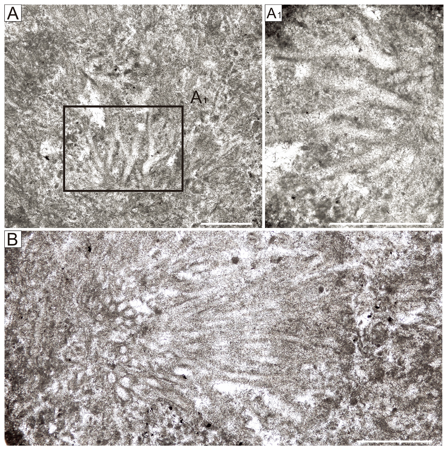
Figure 14(A–B) Botomaella sp. A, Limei section, sample no. BP12-2, longitudinal sections. (A1) Enlargement of part of (A) showing branches in longitudinal section. (B) Overall morphology, Limei section, sample no. BP12-2, longitudinal and cross-sections. Scale bars: (A) 0.2 mm; (A1) and (B) 0.5 mm.
-
Botomaella sp.
Description. Diameters range from 38 to 48 µm; dichotomously branched, branching angles around 35° (Fig. 14A, A1); filaments are long, sometimes up to 5 mm (Fig. 14B).
Remarks. In these specimens, the filaments are larger in diameter prior to branching and smaller after branching, suggesting the presence of main and secondary branches (Fig. 14A). This differs from Ortonella, in which tube diameters remain relatively unchanged after branching. Subradial bundles occur, with branching angles ranging from approximately 25 to 40°. These branching angles appear to be larger than those reported in current species of Botomaella; therefore, we do not assign a species.
-
Genus Epiphyton Bornemann, 1886
(Fig. 15A–D)
-
1967 Paraepiphyton Wray: 41
-
Type species. Epiphyton flabellatum Bornemann, 1886
Diagnosis. Calcified dendritic microfossil, filaments circular, typically micritic, diameter can increase slightly distally, branching often dichotomous at relatively small angles.
Comparison. Korde (1973) described numerous genera broadly similar to Epiphyton, which may include junior synonyms (Riding, 1991b). Nonetheless, despite similarities, several distinct genera can be distinguished (e.g., Gordonophyton, Korilophyton, Tharama, Tubomorphophyton). These have been attributed to the family Epiphytaceae (Korde, 1959, 1973; Chuvashov et al., 1987) and to the Epiphyton Group (Riding, 1991a; Luchinina, 2009).
Affinity. Bornemann (1886) described Epiphyton as a green alga belonging to the Siphonales (Chlorophyta), but its taxonomic attribution remains unresolved. Pia (1927, p. 39) placed Epiphyton in the cyanobacteria. Korde (1959) and Luchinina and Terleev (2008) suggested that Epiphyton is a rhodophyte. Riding and Voronova (1982) suggested that Epiphyton fossils could represent a heterogeneous group. Woo and Chough (2010) provided direct evidence for photosynthesis in Epiphyton fossils from the Middle Cambrian in China. The difficulty of identifying definitive modern analogues, together with the likely heterogeneity of these fossils, has often led to the provisional placement of Epiphyton among calcimicrobes or Microproblematica (Riding, 1991a; Liu et al., 2016a). Min et al. (2019) interpreted Epiphyton as a cyanobacterium based on specimens from the Dengying Formation (Neoproterozoic) at the Lijiagou section in China. However, their material is preserved through phosphatization, which cannot be directly compared with calcified Epiphyton. Ibarra and Sanon (2019) suggested a chlorophyte affinity for Epiphyton.
Species. Epiphyton comprises numerous species, typically based on subtle morphological differences, such as branching angle (Luchinina, 1975; Woo et al., 2008). We distinguish three varieties in our samples.
-
Epiphyton flabellatum Bornemann, 1886
(Fig. 15A, B)
-
1886 Epiphyton flabellatum Bornemann: 18, pl. 1.10.
-
1973 Epiphyton flabellatum Bornemann; Korde, 306, pl. 30.3.
-
2008 Epiphyton flabellatum Bornemann; Woo et al., 59, fig. 5.c.
-
2024 Epiphyton flabellatum Bornemann; Zhu et al., 8, fig. 7.
-
2024 Epiphyton flabellatum Bornemann; Zheng et al., 63 (10), fig. 6A.
-
2025 Epiphyton flabellatum Bornemann; Xiao et al., fig. 10D, E.
Description. Bush-shaped thalli, with radiating micritic branches that are distinct and rounded in transverse section; filament diameter ∼ 50–60 µm; branching is normally dichotomous at 15–20°; rarely segmented.
Remarks. Bifurcating rodlike branches and overall shrub-like form are defining features of Epiphyton flabellatum. Its overall characteristics and dimensions are similar to E. pseudoflexuosum, Epiphyton naturale, and E. tuberculatum. One or more of these may prove to be a synonym of the form described here.
-
Epiphyton parapusillum Korde, 1973
(Fig. 15C)
Description. Thallus hemispheric; outer boundaries of the small bushes are well defined; filament diameter ∼ 50 µm; branching dichotomous at small angles ∼ 10°; filaments lack segmentation.
Remarks. In overall structure and thallus characteristics, this E. parapusillum closely resembles Epiphyton furcatum and Epiphyton fruticosum. It is distinguished by its branching pattern (exhibiting only two to three orders of branching), larger chambers in the fan-shaped sections of the thalli, and exceptionally small size.
-
Epiphyton sp.
(Fig. 15D)
-
2014 Epiphyton sp.; Liu, 72, fig. 4-9A
Description. Filaments densely arranged, creating dendritic radiating clusters up to 4 mm in diameter; individual filaments locally appear to be tubiform with diameter 40–50 µm; branching normally dichotomous at 10–15°; segmentation lacking.
Remarks. Hollow tubes are consistent with Epiphyton sp. reported by Liu et al. (2016a) and can be compared to Tubomorphophyton (see Riding and Voronova, 1982). Scarcity of these fossils in our material (Fig. 15D) makes it difficult to assign this microfossil to a particular species.
-
Genus Renalcis Vologdin, 1932
Type species. Renalcis granosus Vologdin, 1932; Middle Cambrian; Altai Mountains, Russia.
Diagnosis. Calcified, clusters of thick-walled, well-defined, irregularly spherical chambers, forming swollen, hollow aggregates.
Comparison. Nephelostroma Dangéard and Doré, 1957, is regarded as a junior synonym of Renalcis (Reitlinger, 1960).
Affinity. Renalcis has been suggested to be a chroococcalean cyanobacterium (Hofmann, 1975; Pratt and Schlaifer, 1984; Luchinina in Chuvashov et al., 1987; Riding, 1991b; Turner et al., 2000), but its affinities and formation have also long been regarded to be problematic and remain unclear. For example, it has also been suggested to have been produced by precipitation induced by communities of phototrophic and heterotrophic bacteria (Chafetz and Guidry, 1999) or heterotrophic bacteria only (Stephens and Sumner, 2002). Due to a lack of definite present-day analogs, Renalcis and similar genera, such as Izhella, are often broadly regarded to be possible bacterial-calcified Microproblematica (Feng et al., 2010; Liu et al., 2016a, 2017).
-
Renalcis granosus Korde, 1961
(Fig. 15E)
-
1932 Renalcis granosus Vologdin, p. 15, pl. 9.
-
1972 Renalcis sp. Riding and Toomey: 514, pl. 2, figs. 1–3.
-
1973 Renalcis granulatus Korde, p. 112, pl. 2, figs. 2–3.
-
1973 Renalcis erbinatus Korde, p. 114, pl. 1, fig. 4.
-
1990 Renalcis devonicus Bian and Zhou: pl. 1, figs. 2, 5.
-
1995 Renalcis sp. Ye et al.: 15, pl. 8, fig. 6.
-
2009 Renalcis Vologdin; Wang et al.: fig. 2d.
-
2009 Izhella Antropov; Wang et al.: fig. 2e.
-
2011 Renalcis sp. Liu et al.: 501, pl. 5, fig. 6.
-
2016 Renalcis granulatus Liu et al.: 201, fig. 9A.
-
2021 Renalcis granulatus Liu et al.: 18, pl. 9, fig. 7.
Description. Branches of superposed chambers arise from a common base; basal chambers up to 0.55 mm in external diameter. Walls ∼ 50 µm thick and can exhibit poorly developed clefts on their inner surfaces.
Remarks. Relatively large chambers, ranging 200 to 500 µm in external diameter. Recognition of R. granosus is based on morphology, size, and structure, including superimposed chambers, large chamber cavities, and branches arising from a common base.
-
Genus Izhella Antropov, 1955
Type species. Izhella nubiformis Antropov, 1955; Upper Devonian; Russia.
Diagnosis. Subspherical to botryoidal hollow vesicles with thick, micritic walls, aggregated into clusters.
Comparison. Izhella closely resembles Renalcis (Riding 1991a; Feng et al. 2010), and the two often occur intergrown, leading to frequent confusion and/or misidentification in reports (Wray, 1967; Korde, 1973; Wang et al., 2009). The wall of Izhella is characteristically thick with deep, narrow clefts on its inner surface, whereas the wall of Renalcis can often be thinner with poorly developed or no clefts.
-
Izhella nubiformis Antropov, 1955
(Fig. 15F)
-
1955 Izhella nubiformis Antropov: 47.
-
1967 Renalcis turbitus Wray: 46, pl. 11, fig. 1.
-
1983 Renalcis Adams: 330, figs. 1, 2.
-
1973 Renalcis tuberculatus Korde: 113, pl. 3, fig. 1.
-
2011 Izhella sp. Liu et al.: 501, pl. 5, fig. 5.
-
2016 Izhella nubiformis Liu et al.: 201, fig. 9C.
Description. Aggregations of thick-walled micritic chambers. The walls contain deep clefts extending outward from the interior surface through approximately up to two-thirds of the wall thickness. In the longitudinal section, the chambers appear to be lunate; basal chambers up to 0.57 mm in external diameter, with compound lobate cavities ∼ 0.3 mm wide; walls ∼ 0.1 mm thick with clefts up to 60 µm deep.
Remarks. In our specimens, narrow inner clefts extend two-thirds of the way through the wall, matching diagnostic features of the Izhella type species.
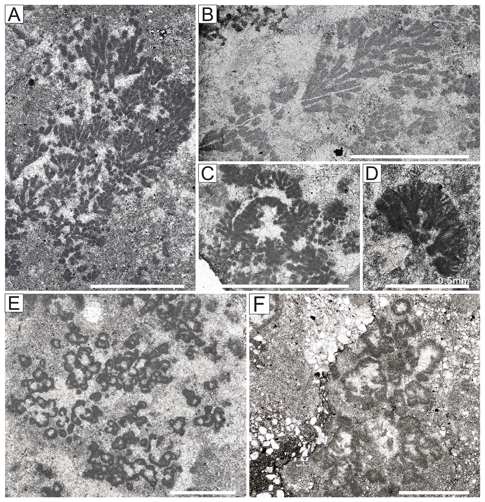
Figure 15(A–D) Epiphyton. (A) Epiphyton flabellatum. Limei section, sample no. LZB-1, longitudinal sections. (B) E. flabellatum. Limei section, sample no. LZB-1, longitudinal sections. (C) Epiphyton parapusillum. Xiachazhuang section, sample no. 6-2, longitudinal and cross-sections. (D) Epiphyton sp., Yutang Section, sample no. 1-11, longitudinal sections. (E) Renalcis granosus, Xiachazhuang section, sample no. 5-1, cross-sections. (F) Izhella nubiformis. Xiachazhuang section, sample no. 6-1, cross-sections. All scale bars equate to 1 mm.
6.1 Global distribution of early Cambrian calcified cyanobacteria
Previous detailed reports of Cambrian calcified cyanobacteria predominantly originated from studies of well-preserved specimens in the Ural Mountains, Siberia, and in North China (Vologdin, 1932, 1937, 1939; Maslov, 1956; Korde, 1961, 1973; Voronova et al., 1969; Luchinina, 1975; Luchinina and Tikhomirova, 1987; Voronova, 1976, 1979; Xiao et al., 2025), with relatively fewer studies in the Americas, Australia, China, Europe, and India, although Epiphyton was initially described from Sardinia (Bornemann, 1886). Detailed distributions are compiled in Table 1.
Table 1Outline of global distribution of principal calcified cyanobacteria and associated calcimicrobe floras during the Cambrian Fortunian–Age 4. Genera in boldface represent new additions from this work.
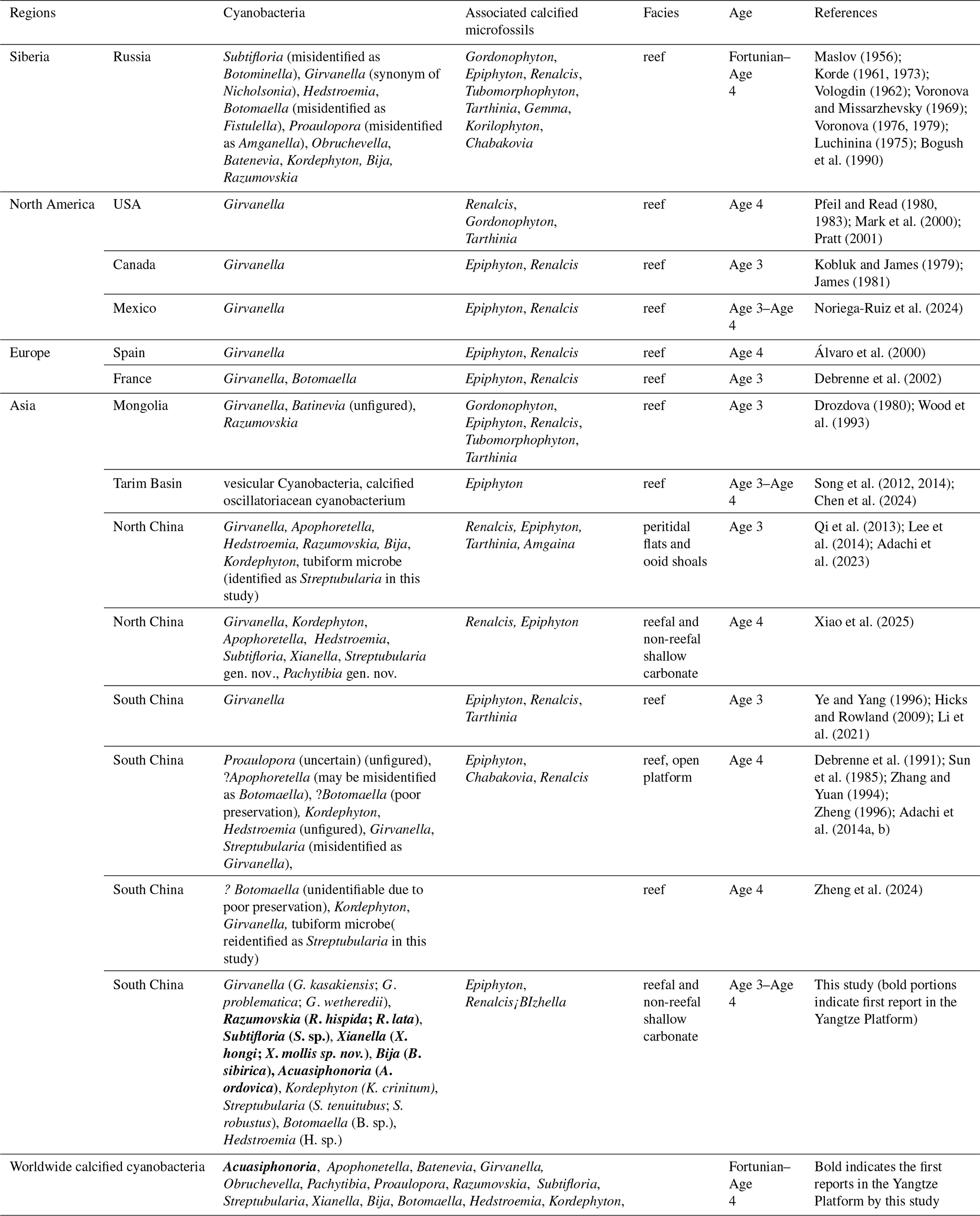
Historically, the Siberian record of calcified cyanobacteria is the most thoroughly studied and has led to major contributions (e.g., Vologdin, 1932; Maslov, 1956; Korde, 1973). Subsequent taxonomic advances regarded Vologdinella Korde, 1957, and Amganella Reitlinger, 1959, as junior synonyms of Proaulopora Vologdin, 1937, and subsumed Botominella Reitlinger, 1959, within Subtifloria Maslov, 1956. Detailed studies (Vologdin, 1932, 1939, 1962; Maslov, 1956; Luchinina, 1975; Korde, 1957, 1973; Voronova, 1976, 1979; Wood et al., 1993; Drozdova, 1980; Kobluk and James, 1979; Riding, 1991a, 2001) have recognized at least 10 genera of early Cambrian calcified microfossils comparable with calcified cyanobacteria: Girvanella, Razumovskia, Subtifloria, Obruchevella, Batenevia, Bija, Botomaella, Hedstroemia, Kordephyton, and Proaulopora (Table 1). In contrast, early Cambrian calcified microfossils documented from North America and Europe are relatively limited in diversity, with only Girvanella and Botomaella (Table 1) commonly being identified (Pfeil and Read, 1980, 1983; Kobluk and James, 1979; James, 1981; Álvaro et al., 2000; Pratt, 2001; Debrenne et al., 2002; Noriega-Ruiz et al., 2024).
Additional occurrences of early Cambrian calcified cyanobacteria and related microfossils have been reported from the North China Block, with genera including Girvanella, Streptubularia, Razumovskia, Subtifloria, Xianella, Apophonetella, Pachytibia, Hedstroemia, Kordephyton, and Bija (Qi et al., 2013; Lee et al., 2014; Adachi et al., 2023; Xiao et al., 2025) (Table 1).
In addition, Girvanella, Kordephyton, Hedstroemia, and Streptubularia have been documented from South China, including the provinces of Hunan, Hubei, and Guizhou (Sun et al., 1985; Zhang and Yuan, 1994; Ye and Yang, 1995; Hicks and Rowland, 2009; Adachi et al., 2014a, b; Li et al., 2021) (Table 1). Among these, Proaulopora and Botomaella cannot be confirmed in the Yangtze Platform from previous studies (Zhang and Yuan, 1994) (lack of figures and/or poor preservation). A study of an archaeocyath reef in the Xiachazhuang section, Hubei Province, reported Botomaella (Zheng et al., 2024); however, re-inspection suggests that these specimens are poorly preserved and may more likely represent Apophoretella (Table 1).
6.2 New discovery of early Cambrian calcified cyanobacteria in South China
Based on the above systematic paleontological studies, 15 species within 10 genera of calcified cyanobacteria and 5 species within 3 genera of Microproblematica are identified in this study from Cambrian Series 2 of South China, including two new species (Streptubularia robustus sp. nov. and Xianella mollis sp. nov.) (Table 1). Thus, our study confirms the presence of four previously reported distinct genera in the early Cambrian of the Yangtze Platform, namely Girvanella, Kordephyton, Hedstroemia, and Streptubularia (Table 1), together with the first documented occurrences of Bija, Botomaella, Acuasiphonoria, Razumovskia, Subtifloria, and Xianella. Recognition of Acuasiphonoria, previously only recognized in Late Ordovician strata, therefore extends its fossil record to the Cambrian.
In addition, previous studies of early Cambrian calcified cyanobacteria have largely focused on reef facies. Our research demonstrates, for the first time, that calcified cyanobacteria also existed in non-reef shallow-marine carbonate facies in South China during this period. The taxonomic composition of the calcimicrobial genera closely follows changes in sedimentary environments. In non-reefal facies, calcimicrobes are very depauperate and typically occur in low-energy hydrodynamic settings. In reef facies, by contrast, calcimicrobe diversity and abundance increase markedly and vary according to the nature of the additional reef builders.
During deposition of the Xiannüdong Formation (Cambrian Series 2, ϵ2x), microbial–archaeocyath reefs developed in South China. Calcified cyanobacteria and problematic calcimicrobes are low in abundance, and the most typical microbial association consists of Epiphyton and Renalcis. The substantial terrigenous clastic input during this period likely influenced the development of calcified cyanobacteria. These microbes either encrust archaeocyaths or occur sporadically in inter-archaeocyath spaces, playing a relatively minor role in reef construction. In contrast, in the Tianheban Formation (ϵ2t), archaeocyaths still serve as framework builders, but the proportion of calcified cyanobacteria is higher. Kordephyton, Streptubularia, and Bija are present and played an important role in reef construction as encrusters and binders. When archaeocyaths became extinct in the Qingxudong Formation (ϵ2q), reefs were subsequently built entirely by calcimicrobes, which show a marked increase in diversity and abundance. Widespread and abundant forms – such as Girvanella, Razumovskia, Xianella, Huayuanella, Kordephyton, and dendritic Epiphyton – reduced water flow velocity, trapped sedimentary particles, and formed the reef framework. Smaller forms (e.g., Girvanella, Razumovskia, Streptubularia) acted as binders, whereas chambered Epiphyton and Renalcis clusters reinforced the framework and occupied interstitial spaces.
6.3 Early Cambrian cyanobacterial calcification episode (CCE) and its implications
During early Cambrian Series 2, calcified cyanobacteria in South China reefal facies gradually evolved from a few relatively simple types, dominated by Oscillatoriaceae, to more diverse assemblages that included Phormidiaceae and Rivulariaceae. This trend may have been driven by ecological competition with archaeocyaths (i.e., an inverse relationship between two reef builders) and by changes in sedimentary environments (i.e., reduction in inputs of terrigenous detrital particles) or both.
Overall, the number of early Cambrian cyanobacterial genera recognized in North China and South China is comparable with that of the Siberian Platform (Table 1). These floras now include 15 genera of calcified cyanobacteria (Table 1): Girvanella, Acuasiphonoria, Batenevia, Razumovskia, Subtifloria, Obruchevella, Streptubularia, Xianella, Pachytibia, Proaulopora, Bija, Botomaella, Hedstroemia, Kordephyton, and Apophoretella. Of these, Acuasiphonoria is the first report of this genus from the Cambrian. In addition, we report two new species (Streptubularia robustus sp. nov. and Xianella mollis sp. nov.). Our findings expand the diversity of early Cambrian cyanobacteria, supporting the observation that high diversity of calcified cyanobacteria in Cambrian Series 2 is a globally recognizable phenomenon.
Cyanobacterial calcification was widespread and occurred across a variety of shallow-marine environments in the Early Cambrian, likely reflecting a significant and widespread change in environmental conditions: a global early Cambrian cyanobacterial calcification episode (CCE). Such episodes have been linked to oceanic and atmospheric conditions that influence marine carbonate precipitation, such as atmospheric pCO2 and oceanic carbonate saturation state (Riding, 1991a, 2006, 2009; Kah and Riding, 2007; Liu et al., 2020). Continued studies of these globally distributed assemblages will provide further new insights into the evolution, biogeographic distribution, and environmental significance of cyanobacterial calcification for understanding early Cambrian marine life and conditions.
Comprehensive systematic taxonomic analysis of reef-associated calcified cyanobacteria from Cambrian Series 2 strata of the Yangtze Platform identified 10 genera, namely Acuasiphonoria, Bija, Botomaella, Girvanella, Hedstroemia, Kordephyton, Razumovskia, Streptubularia, Subtifloria, and Xianella, including two new species (Streptubularia robustus sp. nov. and Xianella mollis sp. nov.). In addition, we recognized three commonly associated genera of uncertain affinity: Epiphyton, Izhella, and Renalcis. This allows recalibration of the inception of the stratigraphic range of Acuasiphonoria, from the Late Ordovician to the Cambrian Series 2. These findings substantially expand the documented diversity of early Cambrian calcified cyanobacteria in non-Siberian terranes and the recognized diversity of their habitats in shallow-marine environments, thereby providing robust evidence to support recognition of the early Cambrian cyanobacterial calcification event. These results improve understanding of Cambrian shallow-marine carbonate communities and sediments and are relevant to the interpretation of factors that influenced seawater chemistry, atmospheric pCO2 levels, and global climate at the inception of the Phanerozoic.
We confirm that no new custom software code was developed or used for the data analysis and interpretation presented in this study. All instrumental data (SEM, EDS, and XRD) were acquired and processed using the standard commercial software packages integrated with the respective instruments (ZEISS Sigma 300 SEM with Bruker Quantax EDS system, and PANalytical Empyrean diffractometer software). These are routine, off-the-shelf tools employed solely for basic data acquisition and standard peak/spectral identification; no custom scripts, macros, or algorithms were written specifically for this research. Therefore, there is no underlying software code that requires deposition in a public repository. All relevant instrument parameters and analytical procedures have been thoroughly described in the Methods section (Sect. 3) of our manuscript, ensuring full reproducibility of the results.
We confirm that all data supporting the findings of this study are presented within the article and its supplementary materials. The primary data include field photographs, polished slab images, thin-section microphotographs, scanning electron microscopy (SEM) images, energy-dispersive X-ray spectroscopy (EDS) spectra, and X-ray diffraction (XRD) patterns, all of which are fully displayed in the main figures (Figs. 1–10) and described in the corresponding figure captions and Methods section (Sect. 3). No raw data were deposited in external public repositories because the key observational and analytical results have been comprehensively documented in the published figures, making the data fully accessible and reproducible from the article itself. Should further details be required, the raw data are available from the corresponding author upon reasonable request.
LL: conceptualization, project administration, supervision, investigation resources, writing (review and editing). JS: preparation, visualization, writing (original draft preparation, review and editing). JZ: visualization, writing (original draft preparation, review and editing). RR: writing (review and editing).
The contact author has declared that none of the authors has any competing interests.
Publisher's note: Copernicus Publications remains neutral with regard to jurisdictional claims made in the text, published maps, institutional affiliations, or any other geographical representation in this paper. The authors bear the ultimate responsibility for providing appropriate place names. Views expressed in the text are those of the authors and do not necessarily reflect the views of the publisher.
We thank Yangge Zhu, Dexiang Gan, Shenhao Xiao, and Weiling Zhuang (NWU) for the assistance in the field work. We thank the editor and the reviewers for the thorough evaluation and constructive suggestions. This study was supported by the National Natural Science Foundation of China (grant nos. 42072127, 42272006, 42472013) and the 111 Project (grant no. D17013).
This research has been supported by the Foundation for Innovative Research Groups of the National Natural Science Foundation of China (grant nos. 42072127, 42272006, 42472013, 42572012) and the 111 Project (grant no. D17013).
This paper was edited by Taniel Danelian and reviewed by three anonymous referees.
Adachi, N., Nakai, T., Ezaki, Y., and Liu, J. B.: Late Early Cambrian archaeocyath reefs in Hubei Province, South China: modes of construction during their period of demise, Facies, 60, 703–717, https://doi.org/10.1007/s10347-013-0376-y, 2014a.
Adachi, N., Ezaki, Y., and Liu, J. B.: The late early Cambrian microbial reefs immediately after the demise of archaeocyathan reefs, Hunan Province, South China, Palaeogeogr. Palaeoclim., 407, 45–55, https://doi.org/10.1016/j.palaeo.2014.04.019, 2014b.
Adachi, N., Natsukawa, H., Ezaki, Y., and Liu, J. B.: Cambrian Series 2 (Stage 4) calcimicrobial reefs in Shandong Province, North China: Calcimicrobial diversity and contribution to reef construction, Palaeogeogr. Palaeoclim., 610, 111351, https://doi.org/10.1016/j.palaeo.2022.111351, 2023.
Adams, A. E.: Lower Carboniferous Renalcis from Cumbria, P. Yorks. Geol. Soc., 44, 327–331, https://doi.org/10.1144/pygs.44.3.327, 1983.
Álvaro, J. J., Vennin, E., Moreno–Eiris, E., and Perejón, A.: Sedimentary patterns across the Lower–Middle Cambrian transition in the Esla nappe (Cantabrian Mountains, northern Spain), Sediment. Geol., 137, 43–61, https://doi.org/10.1016/S0037-0738(00)00132-6, 2000.
Álvaro, J. J., Ezzouhairi, H., Vennin, E., and Ribeiro, M. L.: The Early–Cambrian Boho volcano of the El Graara massif, Morocco: Petrology, geodynamic setting and coeval sedimentation, J. Afr. Earth Sci., 44, 396–410, https://doi.org/10.1016/j.jafrearsci.2005.11.020, 2006.
Antropov, I. A.: Blue-green algae of the Devonian of the central region of the Russian Platform, Kazanskogo Universiteta Uchenye Zapiski, 115, 41–53, 1955 (in Russian).
Arp, G., Reimer, A., and Reitner, J.: Photosynthesis–induced biofilm calcification and calcium concentrations in Phanerozoic oceans, Science, 292, 1701–1704, https://doi.org/10.1126/science.1059046, 2001.
Bian, L. Z. and Zhou, X. P.: A study on calcareous algae from the Upper Ordovician Sanqushan Formation at the border region of Zhejiang and Jiangxi provinces, J. Nanjing Univ. Nat. Sci. Ed. Earth Sci., 2, 1–23, 1990 (in Chinese).
Bogush, O. I., Ivanova, R. M., and Luchinina, V. A.: Izvestkovye vodorosli verkhnego famena i nizhnego karbona Urala i Sibiri, Trudy IGiG SO AN SSSR (Nauka, Novosibirsk), 745, 1–160, 1990 (in Russian).
Bornemann, J. G.: Die Versteinerungen des cambrischen Schichtensystems der Insel Sardinien nebst vergleichenden Untersuchungen über analoge Vorkommnisse aus andern Ländern, Nova Acta Leopold., 51, 1–147, 1886.
Bourque, P. A., Mamet, B., and Roux, A.: Algues siluriennes du Synclinorium de la Baie des Chaleurs, Québec, Canada, Rev. Micropaléontol., 24, 83–126, 1981.
Chafetz, H. S. and Guidry, S. A.: Bacterial shrubs, crystal shrubs, and ray–crystal shrubs: Bacterial vs. abiotic precipitation, Sediment. Geol., 126, 57–74, https://doi.org/10.1016/S0037-0738(99)00032-9, 1999.
Chapman, F.: On the relationship of the genus Girvanella, and its occurrence in the Silurian limestones of Victoria, Report of the Australasian Association for the Advancement of Science, Adelaide, 11, 377–386, 1908.
Chen, D., Yang, B., Song, Y., Guo, C., Zhou, X. Q., Wang, Y. Z., and Li, S. J.: Depositional facies and evolution of sponge-microbial-constructed carbonate platforms in the lower Cambrian (Stage 3), Tarim basin, China: Understanding from anatomy, Mar. Petrol. Geol., 167, 106957, https://doi.org/10.1016/j.marpetgeo.2024.106957, 2024.
Chuvashov, B. I.: Foraminifères et Algues du Dévonien supérieur du versant ouest de l'Oural méridional, in: Foraminifères dévoniens et permiens de l'Oural, Akad. Nauk S. S. S. R. Ural. Filial, 74, 1–153, 1965 (in Russian).
Chuvashov, B. I., Luchinina, V. A., and Shuysky, V. P.: Fossil calcareous algae: morphology, systematics and study methods, Trans. Inst. Geol. Geophys. Sib. Branch USSR Acad. Sci., Novosibirsk, 674, 1–225, 1987 (in Russian).
Conway Morris, S.: The Burgess shale fauna and the Cambrian explosion, Science, 246, 339–346, https://doi.org/10.1126/science.246.4928.339, 1989.
Conway Morris, S.: Ediacaran-like fossils in Cambrian Burgess Shale-type faunas of North America, Paleontology, 36, 593–635, 1993.
Crimes, T. P.: The record of trace fossils across the Proterozoic–Cambrian boundary, in: Origins and Early Evolution of Metazoa, edited by: Lipps, J. H. and Signor, P. W., Plenum Press, New York, 177–202, ISBN 978-1-4899-2429-2, 1992.
Dangéard, L. and Doré, F.: Observations nouvelles sur les Algues et les Stromatolithes du Cambrien de Carteret (Manche), Bull. Soc. Géol. Fr., 7, 1069–1075, https://doi.org/10.2113/gssgfbull.S6-VII.8-9.1069, 1957.
Danielli, H. M. C.: The fossil alga Girvanella Nicholson and Etheridge, Bulletin of the British Museum (Natural History), Geology Series, 35, 79–107, 1981.
Debrenne, F., Gandin, A., and Zhuravlev, A. Y.: Paleoecological and sedimentological remarks on some Lower Cambrian sediments of the Yangtse Platform (China), Bull. Soc. Géol. Fr., 162, 575–583, https://doi.org/10.2113/gssgfbull.162.3.575, 1991.
Debrenne, F., Gandin, A., and Courjault–Radé, P.: Facies and depositional setting of the Lower Cambrian archaeocyath–bearing limestones of southern Montagne Noire (Massif Central, France), Bull. Soc. Géol. Fr., 173, 533–546, https://doi.org/10.2113/173.6.533, 2002.
Dragastan, O.: Review of Tethyan Mesozoic algae of Romania, in: Paleoalgology: Contemporary Research and Applications, edited by: Toomey, D. F. and Nitecki, M. H., Springer-Verlag, Berlin, 101–161, ISBN 0387153128, 1985.
Dragastan, O.: New criteria for the classification of the Porostromata algae, Revista Española de Micropaleontología, 25, 59–89, 1993.
Drosdova, N. A.: Algae in Lower Cambrian organic mounds of western Mongolia, Sovmestnaya Sovetsko-Mongol'skaya Paleontologicheskaya Ekspeditsiya, 18, 137, 1–140, 1980 (in Russian).
Drozdova, N. A.: Algae in Lower Cambrian organogeneous buildups of Western Mongolia, Trans. Joint Soviet Mongol. Palaeontol. Exped., 10, 1–140, 1980 (in Russian).
Elenkin, A. A.: Sinezeleniye vodorosli SSSR. Monografiya presnovodnykh i nazemnykh S'yanofiseaye, obnaruzhennykh v predelakh SSSR. Spetsial'naya (sistematicheskaya) chast', vyp. II. III. Hormogoneae (Geitl.) Elenk. (okonchaniye), Izdate'stvo Akademii Nauk SSSR, Moscow, 985–1908, 1949 (in Russian).
Elicki, O.: Palaeoecological significance of calcimicrobial communities during ramp evolution: An example from the Lower Cambrian of Germany, Facies, 41, 27–39, https://doi.org/10.1007/BF02537458, 1999.
Elliott, G. F.: Algues calcaires codiacées fossiles d'Iraq, nouvelles et peu connues, B. Soc. Geol. Fr., 6, 789–795, 1956.
Elliott, G. F.: Transported algae as indicators of different marine habitats in the English middle Jurassic, Palaeontology, 18, 351–366, 1975.
Erwin, D. H. and Tweedt, S.: Ecological drivers of the Ediacaran–Cambrian diversification of Metazoa, Evol. Ecol., 26, 417–433, https://doi.org/10.1007/s10682-011-9505-7, 2012.
Feng, Q., Gong, Y. M., and Riding, R.: Mid–Late Devonian Calcified Marine Algae and cyanobacteria, South China, J. Paleontol., 84, 569–587, https://doi.org/10.1666/09-142.1, 2010.
Feng, Z. Z., Peng, Y. M., Jin, Z. K., and Liu, Z.: Lithofacies palaeogeography of the Cambrian in South China, J. Palaeogeogr., 3, 1–14, 2001.
Frémy, P. and Dangéard, L.: Sur la position systématique des Girvanelles, Bull. Soc. Linn. Normandie, 8, 101–111, 1935.
Geitler, L.: A. Paschers Die Süsswasserflora von Deutschlands, Ösrerreichs und der Schweiz, Cyanophyceae, Band 12, Gustav Fisher, Jena, 450 pp., 1925.
Gomont, M.: Monographie des Oscillariées (Nostocacées Homocystées), Deuxième partie – Lyngbyées, Annales des Sciences Naturelles, Botanique, 7, 91–264, 1893.
Guilbault, J. P. and Mamet, B. L.: Codiacées (Algues) ordoviciennes des Basses-Terres du Saint-Laurent, Can. J. Earth Sci., 13, 636–660, 1976.
Hicks, M. K. and Rowland, S. M.: Early Cambrian microbial reefs, archaeocyathan inter–reef communities, and associated facies of the Yangtze Platform, Palaeogeogr. Palaeoclim., 281, 137–153, https://doi.org/10.1016/j.palaeo.2009.07.017, 2009.
Høeg, O. A.: Ordovician algae from the Trondheim area, in: The Hovin Group in the Trondheim area, edited by: Kiær, J., Skr. Norske Vidensk.-Akad. Oslo, I. Mat.-Naturvidensk. Kl., 4, 63–96, 1932.
Hofmann, H. J.: Stratiform Precambrian stromatolites, Belcher Islands, Canada: relations between silicified microfossils and microstructure, Am. J. Sci., 275, 1121–1132, https://doi.org/10.2475/ajs.275.10.1121, 1975.
Hu, H. J. and Wei, Y. X.: Freshwater Algae of China: Systematics, Taxonomy and Ecology, Beijing, Science Press, 1023 pp., ISBN 9787030166333, 2006.
Ibarra, Y. and Sanon, S.: A freshwater analog for the production of Epiphyton-like microfossils, Geobiology, 17, 589–602, https://doi.org/10.1111/gbi.12341, 2019.
James, N. P.: Megablocks of calcified algae in the Cow Head Breccia, western Newfoundland: vestiges of a Cambro–Ordovician platform margin, Geol. Soc. Am. Bull., 92, 799–811, https://doi.org/10.1130/0016-7606(1981)92<799:MOCAIT>2.0.CO;2, 1981.
Kobluk, D. R. and James, N. P.: Cavity–dwelling organisms in Lower Cambrian patch reefs from southern Labrador, Lethaia, 12, 193–218, https://doi.org/10.1111/j.1502-3931.1979.tb00998.x, 1979.
Korde, K. B.: New representatives of siphonous algae, Mater. Princ. Palaeontol., 1, 67–75, 1957 (in Russian).
Korde, K. B.: On aspects of fossil blue–green algae, Mater. Princ. Palaeontol., 2, 112–119, 1958 (in Russian).
Korde, K. B.: Morphology and systematic position of representatives of the genus Epiphyton, Reports of USSR Academy of Sciences (NS), 126, 1087–1089, 1959 (in Russian).
Korde, K. B.: Cambrian algae of the southeastern Siberian platform, Trans. Inst. Palaeontol. USSR Acad. Sci., Moscow, 89, 1–147, 1961 (in Russian).
Korde, F. E.: Fresh data on the taxonomy and evolution of early Paleozoic rhodophyceans, Dokl. Akad. Nauk SSSR, Earth Sci. Sect., 166, 197, 1–1441, 1966 (in Russian).
Korde, K. B.: Cambrian algae, Trans. Inst. Palaeontol. USSR Acad. Sci., Moscow, 39, 1–349, 1973 (in Russian).
Kruse, P. D. and Reitner, J. R.: Northern Australian microbial-metazoan reefs after the mid-Cambrian mass extinction, Mem. Assoc. Australas. Paleontol., 45, 31–53, 2014.
Latham, A. and Riding, R. Fossil evidence for the location of the Precambrian/Cambrian boundary in Morocco, Nature, 344, 752–754, 1990.
Lee, J. H. and Riding, R.: Xianella: a new mat–forming calcified cyanobacterium from the Middle–Late Ordovician of North China, Pap. Palaeontol., 2, 439–449, https://doi.org/10.1002/spp2.1048, 2016.
Lee, J. H. and Riding, R.: Stromatolite-rimmed thrombolite columns and domes constructed by microstromatolites, calcimicrobes and sponges in late Cambrian biostromes, Texas, USA, Sedimentology 70, 293–334, https://doi.org/10.1111/sed.13048, 2022.
Lee, J. H., Lee, H. S., Chen, J. T., and Woo, J.: Calcified microbial reefs in Cambrian Series 2, North China Platform: Implications for the evolution of Cambrian calcified microbes, Palaeogeogr. Palaeoclim., 403, 30–42, https://doi.org/10.1016/j.palaeo.2014.03.021, 2014.
Li, G. R. and Cong, P. Y.: The earliest calcified cyanobacteria from the Cambrian Terreneuvian at the western margin of the Yangtze Plate, China, Lethaia, 58, 1–14, https://doi.org/10.18261/let.58.2.3, 2025.
Li, G., Steiner, M., Qian, Y., Zhu, M., and Erdtmann, B. D.: Explosive Radiation of early Cambrian skeletal faunas in South China, in: Originations, Radiations and Biodiversity changes – Evidences from the Chinese Fossil Record, edited by: Rong, J., Fang, Z., Zhou, Z., Zhang, R., Wang, X., and Yuan, X., Science Press, Beijing, 41–57, 831–834, https://doi.org/10.1007/978-1-4615-2522-2_10, 2006.
Li, H., Li, F., Li, X., and Zhang, Y.: Development and collapse of the early Cambrian shallow–water carbonate factories in the Hannan–Micangshan area, South China, Palaeogeogr. Palaeoclim., 583, 110665, https://doi.org/10.1016/j.palaeo.2021.110665, 2021.
Li, J. and He, D. F.: Palaeogeography and tectonic-depositional environment evolution of the Cambrian in Sichuan Basin and adjacent areas, J. Palaeogeogr., 16, 441–460, 2014.
Li, Q., Li, Y., and Kiessling, W.: The first sphinctozoan-bearing reef from an Ordovician back-arc basin, Facies, 61, 1–9, https://doi.org/10.1007/s10347-014-0418-0, 2015.
Li, Z. X., Bogdanova, S., Collins, A., Davidson, A., De Waele, B., Ernst, R., Fitzsimons, I., Fuck, R., Gladkochub, D., Jacobs, J., Karlstrom, K., Lu, S., Natapov, L., Pease, V., Pisarevsky, S., Thrane, K., and Vernikovsky, V.: Assembly, configuration, and break–up history of Rodinia: A synthesis, Precambrian Res., 160, 179–210, https://doi.org/10.1016/j.precamres.2007.04.021, 2008.
Liu, L. J., Wu, Y. S., Yang, H. J., and Jiang, H. X.: Ordovician calcified cyanobacteria and associated microfossils from the Tarim Basin, Northwest China: systematics and significance, J. Syst. Palaeontol., 14, 183–210, https://doi.org/10.1080/14772019.2015.1034448, 2016a.
Liu, L. J., Wu, Y. S., Jiang, H. X., and Yang, W.: Calcified rivulariaceans from the Ordovician of the Tarim Basin, Northwest China, Phanerozoic lagoonal examples, and possible controlling factors, Palaeogeogr. Palaeoclim., 448, 371–381, https://doi.org/10.1016/j.palaeo.2016.02.026, 2016b.
Liu, L. J., Wu, Y. S., Jiang, H. X., and Yang, W.: Paleoenvironmental distribution of Ordovician calcimicrobial associations in the Tarim Basin, Northwest China, Palaios, 32, 462–489, https://doi.org/10.2110/palo.2016.086, 2017.
Liu, L. J., Liang, L. Y., Wu, Y. S., and Jiang, H. X.: Ordovician cyanobacterial calcification: A marine fossil proxy for atmospheric CO2, Earth Planet. Sci. Lett., 530, 115950, https://doi.org/10.1016/j.epsl.2019.115950, 2020.
Liu, L. J., Wu, Y. S., Bao, H. P., and Li, L. X.: Diversity and systematics of Middle–Late Ordovician calcified cyanobacteria and associated microfossils from Ordos Basin, North China, J. Paleontol., 95, 1–25, https://doi.org/10.1017/jpa.2021.23, 2021.
Luchinina, V.: Renalcis and Epiphyton as different stages in the life cycle of calcareous algae, Paleontol. J., 43, 463–468, https://doi.org/10.1134/S0031030109040169, 2009.
Luchinina, V. A.: Palaeoalgological characteristics of the Early Cambrian of the Siberian Platform, Trans. Inst. Geol. Geophys. Sib. Branch USSR Acad. Sci., Novosibirsk, 216, 1–99, 1975 (in Russian).
Luchinina, V. A. and Terleev, A. A.: Peculiarities in the development of Early Cambrian calcareous algae on the Siberian Platform, Acta Micropalaeontologica Sinica, 20, https://doi.org/10.16087/j.cnki.1000-0674.2003.01.005, 2003.
Luchinina, V. A. and Terleev, A. A.: The morphology of the genus Epiphyton Bornemann, Geol. Croat., 61, 105–111, https://doi.org/10.1080/11035899309442257, 2008.
Luchinina, V. A. and Tikhomirova, N. S.: On forms of preservation of Early Cambrian algae and their ultrastructure, in: Calcareous algae and stromatolites, USSR Acad. Sci. Siber. Sec., Inst. Geol. Geophys., Novosibirsk, 12–14, 1987 (in Russian).
Mamet, B. and Roux, A.: Algues dévoniennes et carbonifères de la Téthys occidentale, Rev. Micropaléontol., 18, 134–187, 1975.
Mankiewicz, C.: Proterozoic and Early Cambrian calcareous algae, in: The Proterozoic biosphere: a multidisciplinary study, edited by: Schopf, J. W. and Klein, C., Cambridge University Press, Cambridge, New York, Melbourne, 359–367, https://doi.org/10.1017/cbo9780511601064, 1992.
Maslov, V. P.: The alga Girvanella, its ecology and stratigraphic significance, Bull. Mosk. Obshchestva Ispytatelei Prir., Otd. Geol., 54, 89–100, 1949 (in Russian).
Maslov, V. P.: The fossil calcareous algae of the USSR, Tr. Akad. Nauk SSSR, Geol. Ser., 160, 1–301, 1956 (in Russian).
McMenamin, M. A. S., Debrenne, F., and Zhuravlev, A. Yu.: Early Cambrian Appalachian archaeocyaths: Further age constraints from the fauna of New Jersey and Virginia, U.S.A., Geobios, 33, 693–708, https://doi.org/10.1016/S0016-6995(00)80123-0, 2000.
Mei, C., Riaz, M., Wang, L., Latif, K., and Zhang, R.: Development of Middle Cambrian leiolitic bioherms dominated by calcified microbes: A case study of the Xinji Section (North China Platform), Mar. Micropaleontol., 157, 101858, https://doi.org/10.1016/j.marmicro.2020.101858, 2020.
Min, X., Hua, H., Liu, L. J., and Chen, Z.: Phosphatized Epiphyton from the terminal Neoproterozoic and its significance, Precambrian Res., 331, 105358, https://doi.org/10.1016/j.precamres.2019.105358, 2019.
Min, X., Hua, H., Sun, B., and Chen, Z.: Phosphatised calcified cyanobacteria at the terminal Ediacaran and the earliest Cambrian transition stage: Response to the paleoenvironment, Palaeogeogr. Palaeoclim., 111, 105, https://doi.org/10.1016/j.palaeo.2024.105, 2024.
Mou, C. L., Zhou, K. K., Liang, W., and Ge, X. Y.: Sedimentary environment and petroleum exploration of Early Paleozoic source rocks in the Middle–Upper Yangtze area, Acta Geol. Sin., 85, 1–7, 2011.
Mou, C. L., Liang, W., Zhou, K. K., Ge, X. Y., and Chen, X. W.: Lithofacies palaeogeography of the Early Cambrian (Terreneuvian–Series 2) in the Middle–Upper Yangtze region, Sediment. Geol. Tethyan Geol., 32, 41–53, 2012.
Nicholson, H. A. and Etheridge, R.: A Monograph of the Silurian Fossils of the Girvan District in Ayrshire with Special Reference to Those Contained in the “Gray Collection”, Blackwood, Edinburgh, 341 pp., https://doi.org/10.5962/bhl.title.119722, 1878.
Noriega-Ruiz, H. A., Ochoa-Granillo, J. A., Cuen-Romero, F. J., Valenzuela-Tiznado, J. I., Monreal, R., and Buitrón-Sánchez, B. E.: Cambrian (Fortunian to Jiangshanian) depositional environments of northwestern Mexico: Distribution and facies patterns along the western margin of Laurentia, J. South Am. Earth Sci., 147, 105112, https://doi.org/10.1016/j.jsames.2024.105112, 2024.
Ott, E.: Zwei neue Kalkalgen aus den Cassianer Schichten Südtirols (Oberladin, mittlere Trias), Mitteilungen der Bayerischen Staatssammlung für Paläontologie und historische Geologie, 6, 155–166, 1966.
Pentecost, A. and Riding, R.: Calcification in cyanobacteria, in: Biomineralization in Lower Plants and Animals, edited by: Leadbeater, B. S. C. and Riding, R., Systematics Association Special Volume, 30, 73–90, ISBN 9780198577027, ISBN 0198577028, 1986.
Pia, J.: Thallophyta, in: Handbuch der Paläobotanik, edited by: Hirmer, M., Oldenbourg, München, 31–136, https://doi.org/10.1515/9783486755534-008, 1927.
Pia, J.: Die wichtigsten Kalkalgen des Jungpaläozoikums und ihre geologische Bedeutung, Compte Rendu du 2e Congrès pour l'Avancement des Études de Stratigraphie du Carbonifère, Heerlen, 1935, 2, 765–856, 1937.
Pfeil, R. W. and Read, J. F.: Cambrian carbonate platform margin facies, Shady Dolomite, southwestern Virginia, U.S.A., J. Sediment. Res., 50, 91–116, https://doi.org/10.1306/212F7978-2B24-11D7-8648000102C1865D, 1980.
Pollock, J. B.: Blue–green algae as agents in the deposition of marl in Michigan lakes, Annu. Rep. Mich. Acad. Sci., 20, 247–260, 1918.
Pratt, B. R.: Calcification of cyanobacterial filaments: Girvanella and the origin of lower Paleozoic lime mud, Geology, 29, 763–766, https://doi.org/10.1130/0091-7613(2001)029<0763:COCFGA>2.0.CO;2, 2001.
Pratt, J. W. and Schlaifer, R.: On the Nature and Discovery of Structure, J. Am. Stat. Assoc., 79, 9–21, https://doi.org/10.1080/01621459.1984.10477054, 1984.
Pu, X. C., Zhou, H. D., and Wang, X. L.: Cambrian Lithofacies Palaeogeography and Mineralization in South China, Geological Publishing House, Beijing, 191 pp., ISBN 7116012869, ISBN 9787116012864, 1993.
Qi, Y. A., Dai, M. Y., Chang, Y. G., Wang, M., in Li, D.: Oncoids from the Member 2 of Cambrian Series 3 Mantou Formation in Mianchi area, western Henan Province, Geological Journal of China Universities, 19, 634–641, https://doi.org/10.16108/j.issn1006-7493.2013.04.017, 2013 (in Chinese).
Qian, Y. and Bengtson, S.: Palaeontology and biostratigraphy of the early Cambrian Meishucun stage in Yunnan province, South China, Fossils and Strata, 24, 1–156, 1989 (in Chinese).
Radugin, K. Z. and Stepanova, M. V.: Onitchatykh vodoroslyakh dokembriya severo–zapadnoy chasti Vostochnovo Sayana, in: Materialy po geologii i poleznym iskopaemym Zapadnoy Sibiri, Tomsk. Gos. Univ., Tomsk, 60–64, 1964 (in Russian).
Read, J. F. and Pfeil, R. W.: Fabrics of allochthonous reefal blocks, Shady dolomite (Lower to Middle Cambrian), Virginia Appalachians, J. Sediment. Petrol., 53, 761–778, https://doi.org/10.1306/212F8291-2B24-11D7-8648000102C1865D, 1983.
Reitlinger, E. A.: Atlas mikroskopicheskikh organicheskikh ostatkov i problematiki drevnikh tolshch Sibiri [Atlas of the Microscopic Organic Remains and Problematica from the ancient strata of Siberia], Trudy, Institut geologicheskikh nauk, Akademiya nauk SSR, 25, 1–62, 1959 (in Russian).
Reitlinger, E. A.: Mikroskopicheskie organicheskie ostatki i problematika drevnikh olshch yuga Sibirskoy platformy (Microscopic organic remains and problematica from the ancient strata of the Siberian Platform), in: Doklady sovetskikh geologov, Mezhdunar. Geol. Kongr., XXI Sess., Akad. Nauk SSSR, Moscow, 140–148, 1960 (in Russian, with English abstract)
Riding, R. (Ed.): Calcified cyanobacteria, in: Calcareous Algae and Stromatolites, Springer-Verlag, Berlin, 55–87, https://doi.org/10.1007/978-3-642-52335-9_3, 1991a.
Riding, R. (Ed.): Cambrian calcareous cyanobacteria and algae, in: Calcareous Algae and Stromatolites, Springer-Verlag, Berlin, 305–334, https://doi.org/10.1007/978-3-642-52335-9_16, 1991b.
Riding, R.: Skeletal stromatolites, in: Fossil algae: recent results and developments, edited by: Flügel, E., Springer, Berlin, Heidelberg, New York, 57–60, https://doi.org/10.1007/978-3-642-66516-5, 1977a.
Riding, R.: Systematics of Wetheredella, Lethaia, 10, 94, https://doi.org/10.1111/j.1502-3931.1977.tb00605.x, 1977b.
Riding, R.: Temporal variation in calcification in marine cyanobacteria, J. Geol. Soc., 149, 979–989, https://doi.org/10.1144/gsjgs.149.6.0979, 1992.
Riding, R.: Calcified algae and bacteria, in: Ecology of the Cambrian Radiation, edited by: Zhuravlev, A. Yu. and Riding, R., Columbia University Press, New York, 445–473, ISBN 9780231106122, ISBN 9780231106139, 2001.
Riding, R.: Cyanobacterial calcification, carbon dioxide concentrating mechanisms, and Proterozoic-Cambrian changes in atmospheric composition, Geobiology, 4, 299–316, https://doi.org/10.1111/j.1472-4669.2006.00087.x, 2006a.
Riding, R.: Microbial carbonate abundance compared with fluctuations in metazoan diversity over geological time, Sediment. Geol., 185, 229–238, https://doi.org/10.1016/j.sedgeo.2005.12.015, 2006b.
Riding, R. and Fan, J. S.: Ordovician calcified algae and cyanobacteria, northern Tarim Basin subsurface, China, Palaeontology, 44, 783–810, https://doi.org/10.1111/1475-4983.00201, 2001.
Riding, R. and Toomey, D. F.: The sedimentological role of Epiphyton and Renalcis in Lower Ordovician Mounds, southern Oklahoma, J. Paleontol., 46, 509–519, 1972.
Riding, R. and Voronova, L.: Calcified cyanophytes and the Precambrian–Cambrian transition, Naturwissenschaften, 69, 498–499, https://doi.org/10.1007/BF00396860, 1982.
Riding, R. and Voronova, L.: Assemblages of calcareous algae near the Precambrian/Cambrian boundary in Siberia and Mongolia, Geol. Mag., 121, 205–210, https://doi.org/10.1017/S0016756800028260, 1984.
Riding, R. and Voronova, L.: Morphological Groups and Series in Cambrian Calcareous Algae, in: Paleoalgology: Contemporary Research and Applications, edited by: Toomy, D. F. and Nitecki, M. H., Springer–Verlag, Berlin–Heidelberg, 56–78, https://doi.org/10.1007/978-3-642-70355-3_6, 1985.
Rong, H., Jiao, Y. Q., Wang, Y. B., Wu, L. Q., and Wang, R.: Distribution and geologic significance of Girvanella within the Yijianfang Ordovician reef complexes in the Bachu area, West Tarim Basin, China, Facies, 60, 685–702, https://doi.org/10.1007/s10347-014-0398-0, 2014.
Rothpletz, A.: Die Kalkalgen, Spongiostromen und einige andere Fossilien aus dem Obersilur Gottlands, Sveriges Geologiska Undersökning, Serie Ca, 10, 57 p., 1913.
Shen, C.: Detailed study on the sedimentary environment of the Xiannüdong Formation at the Tangjiahe section, Gucheng Town, Wangcang County, Master Thesis, Southwest Petroleum University, 67 pp., 2015 (in Chinese with English abstract).
Shu, D. G.: Cambrian explosion: Birth of tree of animals, Gondwana Res., 14, 219–240, https://doi.org/10.1016/j.gr.2007.08.004, 2008.
Song, J., Luo, P., Yang, S., and Yang, D.: Evolution characteristics of microbial reef of Lower Cambrian in Sugaiteblak Area, Tarim Basin, Xinjiang Petroleum Geology, 33, 668–671, 2012 (in Chinese with English abstract).
Song, J., Luo, P., Yang, S., Yang, D., Zhou, C., Li, P., and Zhai, X.: Reservoirs of lower Cambrian microbial carbonates, Tarim Basin, NW China, Petrol. Explor. Dev., 41, 449–459, https://doi.org/10.1016/S1876-3804(14)60051-3, 2014.
Stanier, R. Y.: Division I. The Cyanobacteria, in: Bergey's Manual of Determinative Bacteriology, edited by: Buchanan, R. E. and Gibbons, N. E., Williams and Wilkins Co., Baltimore, 22, 1246 pp., ISBN 9780683011173, 1974.
Stephens, N. P. and Sumner, D. Y.: Renalcids as fossilized biofilm clusters, Palaios, 17, 225–236, https://doi.org/10.1669/0883-1351(2002)017<0225:RAFBC>2.0.CO;2, 2002.
Sun, Y. X., Lin, W. Q., and Zhou, Z. D.: Algal fossils, sedimentary environment and their relationship to mineralization in the Early Cambrian Qingxudong Stage of Huayuan, Hunan, J. Chengdu Univ. Technol., 12, 55–63, 1985.
Tang, H. B.: Clotted micrite in Early Ordovician microbial reefs of South China: A case study from the Huanghuachang section, Yichang, China Rev. Earth Sci., 3, 196–206, 2024.
Turner, E. C., James, N. P., and Narbonne, G. M.: Taphonomic control on microstructure in early Neoproterozoic reefal stromatolites and thrombolites, Palaios, 15, 87–111, https://doi.org/10.1669/0883-1351(2000)0152.0.CO;2, 2000.
Valentine, J. W.: On the Origin of Phyla. University of Chicago Press, Chicago, 614 pp., ISBN 9780226845487, ISBN 0226845486, 2004.
Vologdin, A. G.: Archaeocyaths of Siberia, part 2. Cambrian fauna of Altai limestones, Gos. Nauchno-Tekh. Geol. Izdat., Leningrad, 106 pp., 1932 (in Russian).
Vologdin, A. G.: Arkehotsiaty i rezultaty ikh izucheniya v SSSR (Archaeocyatha and the results of their study in the USSR), Probl. Paleontol., 2–3, 453–481, 1937 (in Russian).
Vologdin, A. G.: Archaeocyaths and algae of the Middle Cambrian of the southern Urals, Probl. Paleontol., 5, 210–245, 1939 (in Russian).
Vologdin, A. G.: The oldest algae of the USSR, Reports of USSR Academy of Sciences, Moscow, 656 pp., 1962 (in Russian).
Voronova, L. G.: Calcareous algae of the Precambrian–Cambrian boundary deposits of the Siberian platform, in: Palaeozoic algae and microphytolites, Voronova, L. G. and Radionova, E. P., Inst. Geol. USSR Acad. Sci., Moscow, 3–85, 156–183, 1976 (in Russian).
Voronova, L. G.: Calcareous algae of the Precambrian and Early Cambrian, Bull. Cent. Rech. Explor. Prod. Elf-Aquitaine, 3, 867–871, 1979.
Voronova, L. G.: Algae, in: The Tommotian Stage and the Cambrian Lower Boundary Problem, edited by: Rozanov, A. Yu., Missarzhevskii, V. V., Volkova, N. A., Voronova, L. G., Krylov, I. N., Keller, B. M., Korolyuk, I. K., Lendzion, K., Michniak, R., Pykhova, N. G., and Sidorov, A. D., Academy of Sciences of the U.S.S.R., Geological Institute, Transaction, 206, 189–195, [English edition 1981, Amerind, New Delhi, 221–227], 1969 (in Russian).
Wang, H.: The Sinian System, in: The Geology of China, edited by: Yang, Z., Cheng, Y., and Wang, H., Oxford University Press, New York, 51–63, 1986.
Wang, J. P., Li, Y., Zhang, Y. Y., Yang, H. J., and Huang, Z. B.: Cyanobacterial community from the reef mound of the Lianglitag Formation (Upper Ordovician), Bachu, Xinjiang, NW China, Acta Micropalaeontologica Sinica, 26, 139–147, 2009 (in Chinese with English summary).
Wethered, E.: On the occurrence of the genus Girvanella in oolitic rocks, and remarks on oolitic structure, Q. J. Geol. Soc., 46, 270–283, https://doi.org/10.1144/GSL.JGS.1890.046.01-04.21, 1890.
Woo, B. J. and Chough, A. S. K.: Growth patterns of the Cambrian microbialite: Phototropism and speciation of Epiphyton, Sediment. Geol., 229, 1–8, https://doi.org/10.1016/j.sedgeo.2010.05.006, 2010.
Woo, J., Chough, S., and Han, Z.: Chambers of Epiphyton thalli in microbial buildups, Zhangxia Formation (Middle Cambrian), Shandong Province, China, Palaios, 23, 55–64, https://doi.org/10.2110/palo.2007.p07-004r, 2008.
Wood, A.: The type–species of the genus Girvanella (calcareous algae), Palaeontology, 1, 22–28, 1957.
Wood, R., Zhuravlev, A. Y., and Anaaz, C. T.: The ecology of Lower Cambrian buildups from Zuune Arts, Mongolia: implications for early metazoan reef evolution, Sedimentology, 40, 829–858, https://doi.org/10.1111/j.1365-3091.1993.tb01366.x, 1993.
Wray, J. L.: Upper Devonian calcareous algae from the Canning Basin, Western Australia, Prof. Contrib. Colo. Sch. Mines, 3, 76 pp., 1967.
Wray, J. L.: Calcareous Algae, Elsevier, New York, 185 pp., ISBN 9780444415363, ISBN 9780080868455, 1977.
Xiao, S. H., Liu, L. J., Zheng, J. W., and Wang, R.: Calcified cyanobacteria from Cambrian Series 2–Miaolingian of the North China Plate, Acta Micropalaeontologica Sinica, 42, 234–257, https://doi.org/10.16087/j.cnki.1000-0674.20250924.001, 2025 (in Chinese).
Ye, J. and Yang, Y. Y.: Ordovician reefs in the Southwestern Margin of the Ordos Basin, Geological Publishing House, Beijing, 67 pp., 1995 (in Chinese).
Ye, J., Yang, Y. Y., Xu, A. D., and Li, W. H.: Early Cambrian reefs in southern Shaanxi, in: Chinese Bioreefs and Petroleum, Palaeozoic Bioreefs, edited by: Fan, J. S., China Ocean Press, Beijing, 18–28, ISBN 9787502738570, 1996 (in Chinese).
Zeng, K.: Characteristics and geological significance of mixed sedimentation in the Cambrian Series 2 Xiannüdong Formation, Northern Sichuan Basin, Master Thesis, Southwest Petroleum University, https://doi.org/10.27420/d.cnki.gxsyc.2020.000379, 2020.
Zhang, H., Dai, M., Qi, Y., and Wu, Y. S.: Girvanella fossils from the Phanerozoic: Distribution, evolution and controlling factors, J. Palaeogeogr., 13, 924–938, 2024.
Zhang, J. M. and Yuan, K. X.: Archaeocyathan reef mounds and their diagenesis of the Lower Cambrian Tianheban Formation at Wangjiaping, Yichang, Hubei, China. J. Geol., 29, 236–245, 1994 (in Chinese).
Zhang, M. L., Xie, Z. Y., Li, X. Z., Yang, Y., and Zhang, X.: Characteristics of Cambrian lithofacies palaeogeography in the Sichuan Basin, Acta Sedimentol. Sin., 28, 128–139, 2010.
Zhang, X., Shu, D., Han, J., and Liu, J.: Triggers for the Cambrian explosion: Hypotheses and problems, Gondwana Res., 25, 896–909, https://doi.org/10.1016/j.gr.2013.06.001, 2014.
Zhang, Y. Y., Li, Q. J., Li, Y., and Yang, Y.: Cambrian to Lower Ordovician reefs on the Yangtze Platform, South China Block, and their controlling factors, Facies, 62, 17, https://doi.org/10.1007/s10347-016-0473-9, 2016.
Zheng, J. W., Zhu, Y. G., Liu, L. J., Shen, J. W., Wang, R., Zhuang, W. L.: Microbial–archaeocyathan reefs in the Cambrian Stage 4 Tianheban Formation at the Xiachazhuang section, Yichang, Hubei Province, and their palaeoecological significance, Acta Palaeontologica Sinica, 63, 310–329, https://doi.org/10.19800/j.cnki.aps.2024001, 2024 (in Chinese).
Zheng, R. C.: Early Cambrian algal reefs in the Yutang area, Huayuan County, western Hunan, in: Chinese Bioreefs and Petroleum, Palaeozoic reefs, edited by: Fan, J. S., China Ocean Press, Beijing, 29–38, ISBN 9787502738570, 1996 (in Chinese).
Zhu, M. Y., Sun, Z. X., Yang, A. H., Zhao, F. C., and Lu, Y.: Division and correlation of Cambrian rock strata in China, J. Stratigr., 45, 223–249, 2021.
Zhu, Y. G., Liu, L. J., Jia, L. Q., Li, L. X., and Zheng, J. W.: Archaeocyathan fossils from the Cambrian Series 2 Xiannüdong Formation, Mianxian County, southern Shaanxi Province, and their biostratigraphic significance, J. Stratigr., 48, 237–252, https://doi.org/10.19839/j.cnki.dcxzz.2024.0021, 2024.
- Abstract
- Introduction
- Geological setting and outcrop sections
- Material and methods
- Systematic paleontology
- Calcified Microproblematica
- Discussion
- Conclusions
- Code availability
- Data availability
- Author contributions
- Competing interests
- Disclaimer
- Acknowledgements
- Financial support
- Review statement
- References
- Abstract
- Introduction
- Geological setting and outcrop sections
- Material and methods
- Systematic paleontology
- Calcified Microproblematica
- Discussion
- Conclusions
- Code availability
- Data availability
- Author contributions
- Competing interests
- Disclaimer
- Acknowledgements
- Financial support
- Review statement
- References