
the Creative Commons Attribution 4.0 License.
the Creative Commons Attribution 4.0 License.
 | 27 Aug 2026
Taxonomic reassessment and palaeobiogeographical patterns of some Miogypsinidae species, larger benthic foraminifera
Juan Carlos Braga
Shunichi Kinoshita
Johannes Pignatti
Yasufumi Iryu
The Miogypsinidae comprise orbitoidal larger benthic foraminifera with generally eccentric position of the nepionic stage and a fan of equatorial chamberlets; the members of this family are widely used in biostratigraphy of upper Oligocene–middle Miocene deposits. Here, a taxonomic review of the genus Miogypsinella Hanzawa, 1940 (type species: M. borodinensis Hanzawa, 1940) and Miogypsinoides lateralis Hanzawa, 1940 is presented based on the reassessment of the type specimens. The palaeobiogeographical patterns of these taxa are discussed based on a compilation of the geographical and stratigraphical ranges of all their reported occurrences including illustrations. The computed tomography (CT)-scan analysis of the type material of M. borodinensis, from the Chattian of the central Indo-Pacific (Kitadaito Jima), indicates that it is a valid species different from Miogypsinoides complanatus. Two Miogypsinella species (M. borodinensis, M. ubaghsi) are widespread in the Chattian–Aquitanian of the Indo-Pacific region and one species (M. bermudezi) in the Aquitanian of the eastern Pacific (Mexico). Miogypsinella appeared in the Chattian in the central Indo-Pacific, reached Central America, and disappeared in the Aquitanian in both areas. The American Oligocene faunas of larger benthic foraminifera were, therefore, not distinctly isolated as previously proposed. Miogypsinoides lateralis is a clearly distinct species that is an exception to the presumed morphological trend within the Miogypsinoides species lineage. Miogypsinoides lateralis possesses morphometric values comparable to those of the Burdigalian–Langhian M. indica but occurring earlier, in the Aquitanian. The latest Aquitanian initiation of the Indonesian Seaway restriction and widespread drowning of former carbonate platforms likely brought about the extinction of Miogypsinella borodinensis, M. bermudezi, M. ubaghsi, and Miogypsinoides lateralis.
- Article
(17782 KB) - Full-text XML
- BibTeX
- EndNote


Miogypsinids (Miogypsinidae Vaughan, 1928 family) constitute a group of orbitoidal larger benthic foraminifera (LBF) with hyaline–lamellar perforated shell, spiral-arranged nepionic chambers with later equatorial chamberlets, and spiral and intraseptal canals (e.g. de Bock, 1976; Chaproniere, 1984; Loeblich and Tappan, 1987; Drooger, 1993). This group differs from other orbitoidal forms in having a generally eccentric position of the nepionic stage, located near the apex of the shell, and the development of equatorial chamberlets in one sector only (e.g. de Bock, 1976; Drooger, 1993). A total of 5 miogypsinid genera were accepted by Loeblich and Tappan (1987), 7 by BouDagher-Fadel and Price (2013), 8 by BouDagher-Fadel (2018), and 14 by Hayward et al. (2021). Miogypsinoides Yabe and Hanzawa, 1928 and Miogypsina Sacco, 1893 have so far been the most studied taxa, recorded from the western Tethys through Indo-Pacific areas and to Central America (Drooger, 1952, 1953, 1963; Drooger and Socin, 1959; Raju, 1974; BouDagher-Fadel and Price, 2013).
Miogypsinids have been used in biostratigraphy of Oligocene–middle Miocene deposits in both the western Tethys and in Indo-Pacific areas (e.g. Raju, 1974; Adams, 1984; Drooger and Laagland, 1986; Cahuzac and Poignant, 1997; Lunt and Allan, 2004; BouDagher-Fadel, 2018).
Based on Indonesian material, Tan (1936a, b, 1937a, b) found that the number of the nepionic chambers (hereafter, X value; i.e. nepionic stage) progressively decreased in the populations through time, with primitive forms (i.e. Miogypsinoides) having a longer nepionic stage, a trend named nepionic acceleration. This temporal morphological change was applied together with other shell characters in morphometric taxonomy with biostratigraphical aims, which has been historically prevailing in miogypsinid studies (e.g. Drooger, 1963, 1993; Raju, 1974; Schiavinotto, 1985; Özcan et al., 2009; Schiavinotto and Benedetti, 2021). In orbitoidal or radial foraminifera, the shell morphology may be very different at the beginning and end of the lineages. For these groups, including miogypsinids, Drooger (1993) proposed to split the individual lineages into segments based on morphometric data. These segments, which have been called species, are defined by a morphometric range (Pignatti, 1998). This species concept circumscribes artificial units with well-defined but arbitrarily chosen morphometric limits (e.g. Drooger, 1993; Özcan et al., 2007, 2009; Less, 1987).
The origin of the family is uncertain, and the geographical location of the ancestral forms is still debated. Mexico, south-western France, and Spain have been proposed as possible centres of origination of an Oligocene ancestor (Salmeron, 1972; Cahuzac and Poignant, 1987; Laagland, 1990). Two miogypsinid species, Miogypsinella borodinensis Hanzawa, 1940 (type species of the genus) and Miogypsinella bermudezi (Drooger, 1951), have alternatively been considered as possible ancestors of the family (Drooger, 1951, 1963; BouDagher-Fadel and Price, 2013). The hypotheses about the primitive forms were based on morphometric data (e.g. proloculus diameter, length of the nepionic stage), generally comparing them to those of Miogypsinoides complanatus (Schlumberger, 1900), considered by some authors to be the oldest species of the family (e.g. Drooger, 1993; BouDagher-Fadel, 2018). Both hypotheses, however, were deeply influenced by their unclear taxonomic position.
The shell architecture and systematic position of Miogypsinella have been repeatedly discussed in the literature, and it has been often placed in or considered a synonym of the genus Miogypsinoides (e.g. Drooger, 1952; Loeblich and Tappan, 1987; Sirel and Işık, 2011; Matsumaru et al., 2010; Gedik, 2018; Mitchell, 2026). According to Drooger (1993), the frequent changes in species names and concepts and the lack of morphometric details make it difficult to evaluate various reports of Miogypsinella from Pacific areas (e.g. Cole, 1954, 1969). Uncertainties stem from conflicting descriptions of Miogypsinella borodinensis and related species, in many cases incomplete or incorrect (e.g. Sirel and Işık, 2011; Matsumaru et al., 2010; Gedik, 2018), and from different interpretations of some shell structures (and the terminology applied to them). Rigorous descriptions of shell structures, besides their taxonomic value, are important to understand the functional morphology of the test and the relationships of shell architecture to cytoplasmic differentiation (Hottinger and Dreher, 1974; Hottinger, 1978; Hottinger and Leutenegger, 1980). A detailed morphological analysis and taxonomic assessment of the Miogypsinella species are, therefore, needed to circumscribe them, to narrow down their biostratigraphical range, and to discuss their possible phylogenetic relationships.
In the collection of Hanzawa (1940), in addition to Miogypsinella borodinensis, a second miogypsinid species is present: Miogypsinoides lateralis Hanzawa, 1940. Due to its peculiar nepionic stage characterized by a low number of chambers, it has been mixed up with Miogypsinoides species of Oligocene faunas from the western Tethys (Mediterranean) and the Indo-Pacific, and it has only been reported from the central Indo-Pacific (e.g. Hanzawa, 1957, 1962; Cole, 1957b, 1969). The species was later synonymized with Miogypsinoides dehaartii, hampering its use in biostratigraphical schemes (Lunt and Luan, 2022).
This study presents direct observations of shell structure and architecture of two miogypsinid species, Miogypsinella borodinensis and Miogypsinoides lateralis, as well as their systematic implications. The shell characters were observed by computed tomography (CT) scans. This advanced technology allowed us a detailed examination of shell characters, leading to a better understanding of species delimitation and evolutionary relationships. This morphological analysis permits (1) understanding the species taxonomy, (2) setting speciation and extinction events of Miogypsinella species, and (3) tracing the Oligocene–early Miocene biostratigraphical and palaeobiogeographical scenarios for these miogypsinids in the western Tethyan and Indo-Pacific areas. Three species of Miogypsinella can be separated by the proloculus diameter, the number of nepionic chambers, the inclination of the planispiral whorl with respect to the equatorial plane, and the arc length of nepionic spiral from embryonic chambers to the apical point of test.
This study was carried out on material preserved both as thin sections and isolated specimens in the collection of Hanzawa (1940) deposited in the Tohoku University Museum (Sendai, Japan). Oriented sections of LBF specimens in the collection of Hanzawa (1940) from Kitadaito Jima were housed at the Institute of Geology and Paleontology (IGPS), Faculty of Science (currently Department of Earth Science, Graduate School of Science).
The structural and morphological terms are those used by de Bock (1976), Drooger (1993), Hottinger et al. (1991), and Hottinger (2006). The suprageneric classification follows Loeblich and Tappan (1987). The descriptions of the analysed Miogypsinella species are ordered according to their stratigraphical appearance. References to published species records include only those in which structural diagnostic features were described and illustrated.
The isolated specimens were micro-CT-scanned at the Centro de Instrumentación Científica, Universidad de Granada, Spain. The micro-CT scanning system Zeiss Xradia 510 Versa with granite-based vibratory insulation was used. The source voltage used was 30–160 kV, with 2000×2000 pixel noise detector suppression at full charge. Non-destructive three-dimensional advanced image solutions were formed with high-contrast images and submicron resolution (700 nm). The true spatial resolution was 0.7 µm and 70 nm voxel size. The micro-computed tomographic scanning 3D models were rendered using Molcer Plus (Ver. 1.6) with the shell removed to highlight the volumes occupied by protoplasm within the shell and the arrangement and communications among chambers.
-
Superfamily Rotalioidea Ehrenberg, 1839
-
Family Miogypsinidae Vaughan, 1928
-
Genus Miogypsinella Hanzawa, 1940
-
Type species. Miogypsinella borodinensis Hanzawa, 1940, pp. 779–780, pl. 39, figs. 1–9.
Diagnosis. Test flat and concavo-convex, rarely conical; embryo near test apex consisting of a large sub-spheroidal protoconch and reniform deuteroconch separated by an imperforated wall and both surrounded by a perforated wall; protoconchal stolon lies in the opposite side of the deuteroconchal aperture; row of peri-embryonic chambers in a single spiral; equatorial chamberlets asymmetrical and ogival; apertural lip; septal flap; intraseptal interlocular space occurring around each embryonic, nepionic, and equatorial chamber and chamberlet; cover on intraseptal interlocular space; consecutive toothplates interconnected in adaxial position producing a primary spiral canal; intraseptal canal system shows vertical branches which issue from the spiral canal and extend to ventral side only; subsutural canals; very thin single-layered bilamellar outer shell wall in the equatorial chambers and chamberlets.
Remarks. Miogypsinella differs from Miogypsinoides in having a cover on intraseptal interlocular space, an apertural lip, and subsutural canals (Table 1). The higher number of planispiral nepionic chambers (Drooger and Raju, 1973; de Bock, 1976; Matsumaru, 2012), a broader flange becoming laterally heavily thickened, and a weakly trochospiral initial coil (Hanzawa, 1962, figs. 1–11; Loeblich and Tappan, 1987; BouDagher-Fadel et al., 2000; Matsumaru, 2012; BouDagher-Fadel and Price, 2013; BouDagher-Fadel and Wilson, 2000) have been used so far as distinctive shell characters to separate Miogypsinoides from Miogypsinella. These characters, however, are not supported by morphological evidence. The use of a “stronger predominance of pillars and pustules” to circumscribe Miogypsinoides, implying that Miogypsinella is its younger synonym (Cole in Cushman, 1948, p. 376; Drooger, 1953, p. 120; Drooger, 1993, p. 81), is not based on distinctive characters at the genus level and, therefore, cannot be accepted.
Table 1Comparison of diagnostic shell characteristics of Miogypsinella and Miogypsinoides. Based on data from 1 Hanzawa (1940), Drooger (1952, 1993), BouDagher-Fadel and Price (2013), and this study. 2 de Bock (1976), Loeblich and Tappan (1987), Drooger (1952, 1963, 1993), BouDagher-Fadel and Price (2013), and this study. Abbreviations: co, cover on intraseptal interlocular space; iis, intraseptal interlocular space; li, apertural lip; ssc, subsutural canals; sf, septal flap; spc, primary spiral canal; ssc, subsutural canals; tlw, thickness of the lateral walls; tp, toothplate; vcs, vertical canal system.

Drooger (1953) stated that Miogypsinoides differs from Miogypsinella in having side walls of distinctly lamellar structure, whereas the latter shows “dominating vertical structures” (Hanzawa, 1940; pillars and pustules in Drooger, 1993; BouDagher-Fadel and Price, 2013) in these lateral walls. After recognizing that these characters do not provide a sharp demarcation between the two taxa, he considered Miogypsinella to be a junior synonym of Miogypsinoides, including in this genus all species without lateral chamberlets (Drooger, 1953, p. 120, 122; Drooger, 1993, p. 81). According to this author, the occurrence of “some cavities of various kinds in the side walls” (i.e. intraseptal interlocular space) and nepionic characters (i.e. trochoid or planispiral whorl) may be regarded as diagnostic at species level (Drooger, 1953, p. 120).
Sirel and Işık (2011) and Gedik (2018) distinguished Miogypsinella from Miogypsinoides in having the “early rotaloid stage with pillars”. Some Miogypsinoides species were transferred to Miogypsinella without any taxonomic comparisons and remarks (Sirel, 2010; Sirel and Işık, 2011; Sirel and Gedik, 2011; Gedik, 2018). Similar taxonomic misconceptions of Miogypsinoides complanatus/Miogypsinella complanata (Schlumberger) (e.g. Sirel and Işık, 2011; Matsumaru et al., 2010; Gedik, 2018) have caused confusion in the stratigraphical range of this species as well as in its palaeobiogeographical distribution.
Hanzawa (1957, p. 86) considered Miogypsinella to be a junior synonym of Miogypsinoides because both genera show nepionic chambers trochospirally arranged (“asymmetrical between the ventral and dorsal sides”). As remarked above, neither this arrangement nor the mere number of nepionic chambers (de Bock, 1976, p. 59) can be used to separate the two genera.
Tan (1936a, p. 51) established Conomiogypsinoides providing two illustrations from thin-sectioned material: a paraxial section and an oblique equatorial section. These specimens show a heavily thickened wall with a vertical canal system on both sides of the shell, suggesting a possible ascription to Miogypsinoides (Hanzawa, 1940, p. 775; Hanzawa, 1962, p. 153).
The World Register of Marine Species (Hayward et al., 2021) reports Miogypsinella Hanzawa, 1940 as a subjective junior synonym of Miogypsinoides Yabe and Hanzawa, 1928 (opinion of Loeblich and Tappan, 1987), and lists seven Miogypsinella species (as Miogypsinoides): M. borodinensis Hanzawa, 1940; M. boninensis Matsumaru, 1996; M. bornea BouDagher-Fadel and Price, 2013; M. cyprea BouDagher-Fadel and Price, 2013, M. elongata BouDagher-Fadel and Price, 2010; M. sanjosensis Hanzawa, 1940; M. ubaghsi Tan, 1936a. Miogypsinella akcadagensis (Gedik and Sirel) and Miogypsinella bermudezi (Drooger) are not listed as such in Hayward et al. (2021).
Miogypsinella boninensis Matsumaru, 1996 (see also Sharaf et al., 2005, 2014), Miogypsinella bornea BouDagher-Fadel and Price, 2013, Miogypsinella cyprea BouDagher-Fadel and Price, 2013, Miogypsinella elongata BouDagher-Fadel and Price, 2010, and Miogypsinella matsumaria BouDagher-Fadel and Price, 2010 have not been sufficiently described and illustrated to assess their status as separate species. In particular, no key information about the apertural lip, spiral canal, subsutural canal, and vertical canal system is given in their descriptions. All these species are characterized by a thickened lateral shell wall, which reasonably allows their assignment to Miogypsinoides (de Bock, 1976; BouDagher-Fadel and Price, 2013; Table 1). Novandaru et al. (2025, fig. 5l–o) identified Miogypsinella bornea from the Chattian of West Java. The proposed shell structures do not clearly distinguish Miogypsinella and Miogypsinoides (Novandaru et al., 2025, table 1).
Miogypsinella akcadagensis (Gedik and Sirel) was first ascribed to Miogypsinoides by Gedik and Sirel (2009) and later placed in Miogypsinella by Gedik (2018). This species shows a thickened lateral wall, suggesting that it belongs to Miogypsinoides rather than to Miogypsinella (Table 1). This conclusion is supported by later records of this species and its illustrations (Gedik and Sirel, 2009; Sirel and Gedik, 2011; Gedik, 2014, 2015, 2018). Miogypsinella bermudezi (Drooger) is described and discussed in the following chapter.
Gedik (2018) proposed that Miogypsinoides (Miogypsinoides) complanatus (Schlumberger) forma bantamensis Tan, 1936a, Miogypsinoides bantamensis, and Miogypsinoides bermudezi Drooger, 1951 are younger synonyms of Miogypsinella borodinensis based on the number of nepionic (spiral) chambers. This synonymy cannot be accepted due to the different shell characteristics of Miogypsinoides and Miogypsinella.
The Miogypsinella species dealt with herein are exclusively fossil (M. borodinensis, M. bermudezi, and M. ubaghsi; Table 3). The proloculus diameter, the number of nepionic chambers, the inclination of the planispiral whorl in relation to the equatorial chamber plane, and the arc length of the nepionic spiral starting from embryonic chambers and ending at the apical point of test (i.e. clockwise spiral; hereinafter, angle γ; Amato and Drooger, 1969) are reliable characters to distinguish the species of this genus (Table 2).
Table 2Comparison of diagnostic shell characteristics of Miogypsinella species and their stratigraphical distribution. Species are listed according to their first appearance reported in the literature. Based on data from 1 Barker and Grimsdale (1937), Hanzawa (1940, 1957, 1965), and this study. 2 Drooger (1951, 1963, 1993), Akers and Drooger (1957), and Hanzawa (1957). 3 Tan (1936a), Cole (1954, 1957b), Hanzawa (1957, 1965), and Mohiuddin et al. (2000). Abbreviations: angle γ, arc length of nepionic spirals starting from embryonic chambers and ending at the apical point of test; L, length (max); prol, proloculus diameter; pwd, planispiral whorl direction degree; nc, number of nepionic chambers.

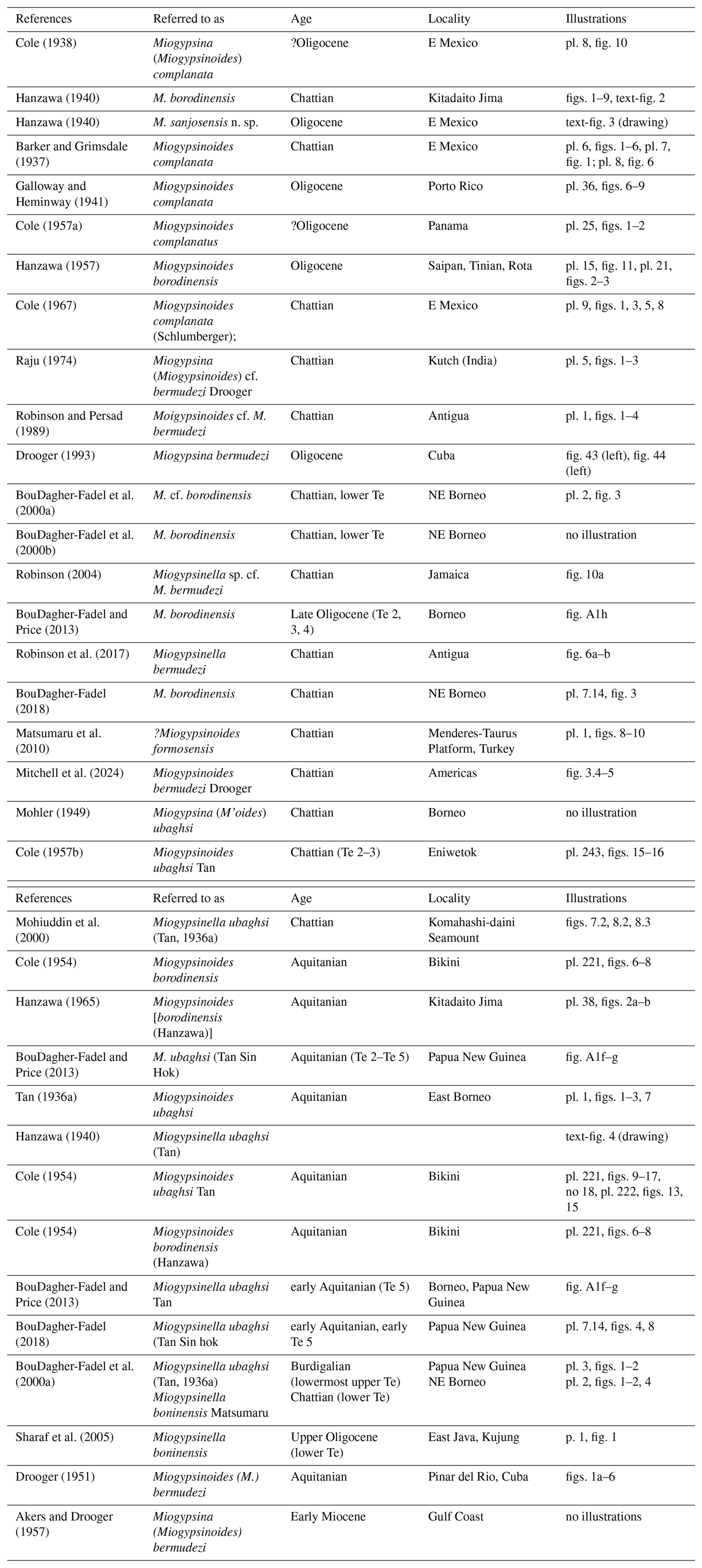
Table 3Stratigraphical and geographical distribution of Miogypsinella borodinensis Hanzawa, Miogypsinella bermudezi (Drooger), and Miogypsinella ubaghsi (Tan).
-
Miogypsinella borodinensis Hanzawa, 1940
Figures 1–5. -
non 1924 Miogypsina complanata Schlumberger; Silvestri, pl. 1, fig. 19.
-
? 1937 Miogypsinoides complanata; Barker and Grimsdale, pl. 5, fig. 6.
-
1937 Miogypsinoides complanata; Barker and Grimsdale, pl. 6, figs. 1–6, pl. 7, fig. 1; pl. 8, fig. 6.
-
1938 Miogypsinella Hanzawa, pp. 387–389.
-
1938 Miogypsina (Miogypsinoides) complanata Schlumberger; Cole, pl. 8, fig. 10.
-
v. 1940 Miogypsinella borodinensis n. sp., Hanzawa, pp. 779–780, pl. 39, figs. 1–9, text-fig. 2.
-
1940 Miogypsinella sanjosensis n. sp., Hanzawa, text-fig. 3.
-
1941 Miogypsinoides complanata (Schlumberger); Galloway and Heminway, pl. 36, figs. 6–9.
-
1951 Miogypsina (Miogypsinella) Drooger, p. 364 (nom. transl.).
-
? 1954 Miogypsinoides borodinensis (Hanzawa); Cole, pp. 600–601, pl. 221, figs. 6–8.
-
? 1957a Miogypsinoides complanatus (Schlumberger); Cole, pl. 25, figs. 1–2
-
1957 Miogypsinoides borodinensis (Hanzawa); Hanzawa, pp. 91–92, pl. 15, fig. 11; pl. 21, figs. 2–3.
-
1962 Miogypsinoides complanatus (Schlumberger); Hanzawa, pl. 7, fig. 11.
-
1962 Miogypsinoides borodinensis (Hanzawa); Hanzawa, pl. 7, fig. 15.
-
1965 Miogypsinoides [borodinensis (Hanzawa)]; Hanzawa, pl. 38, fig. 2a–b.
-
1967 Miogypsinoides complanata (Schlumberger); Cole, pl. 9, figs. 1, 3, 5, 8.
-
1974 Miogypsina (Miogypsinoides) cf. bermudezi Drooger; Raju, p. 77, pl. 1, figs. –5 (drawings); ? pl. 3, figs. 1–2 (outer views); pl. 5, figs. 1-3.
-
? 1984 Miogypsinoides borodinensis Hanzawa; Cahuzac, pl. 1, fig. 5.
-
1988 Miogypsinella Hanzawa; Loeblich and Tappan, p. 680.
-
1989 Miogypsinoides cf. M. bermudezi Drooger; Robinson and Persad, pl. 1, figs. 1–4.
-
? 2000b Miogypsinella cf. borodinensis Hanzawa; BouDagher-Fadel et al., p. 14, pl. 2, fig. 3.
-
2004 Miogypsinella sp. cf. M. bermudezi; Robinson, fig. 10a.
-
? 2010 Miogypsinoides formosensis; Matsumaru et al., pl. 1, figs. 8–10.
-
non 2010 Miogypsinella complanata (Schlumberger); Matsumaru et al., pl. 1, figs. 5–7.
-
2013 Miogypsinella ubaghsi Tan; BouDagher-Fadel and Price, p. 198, fig. A1f–g.
-
? 2013 Miogypsinella borodinensis Hanzawa; BouDagher-Fadel and Price, p. 197, fig. A1h.
-
non 2015 Miogypsinella borodinensis Hanzawa; Sirel, pl. 42, fig. 1; pl. 52, figs. 1–26; pl. 53, fig. 1.
-
2017 Miogypsinella bermudezi Akers and Drooger; Robinson et al., fig. 6a–b.
-
? 2018 Miogypsinella borodinensis Hanzawa; BouDagher-Fadel, pl. 6.30, fig. 3.
-
? 2023 Miogypsinella; Alvarado Sierra et al., fig. 9X.
-
2024 Miogypsinoides bermudezi Drooger; Mitchell et al., fig. 3.4–5.
Type reference and figures. Miogypsinella borodinensis Hanzawa, 1940, pp. 779–780, pl. 39, figs. 1–9 (figs. 1–4, isolated specimens), text-fig. 2 (hand drawing illustrating the specimen of pl. 39, figs. 8–9).
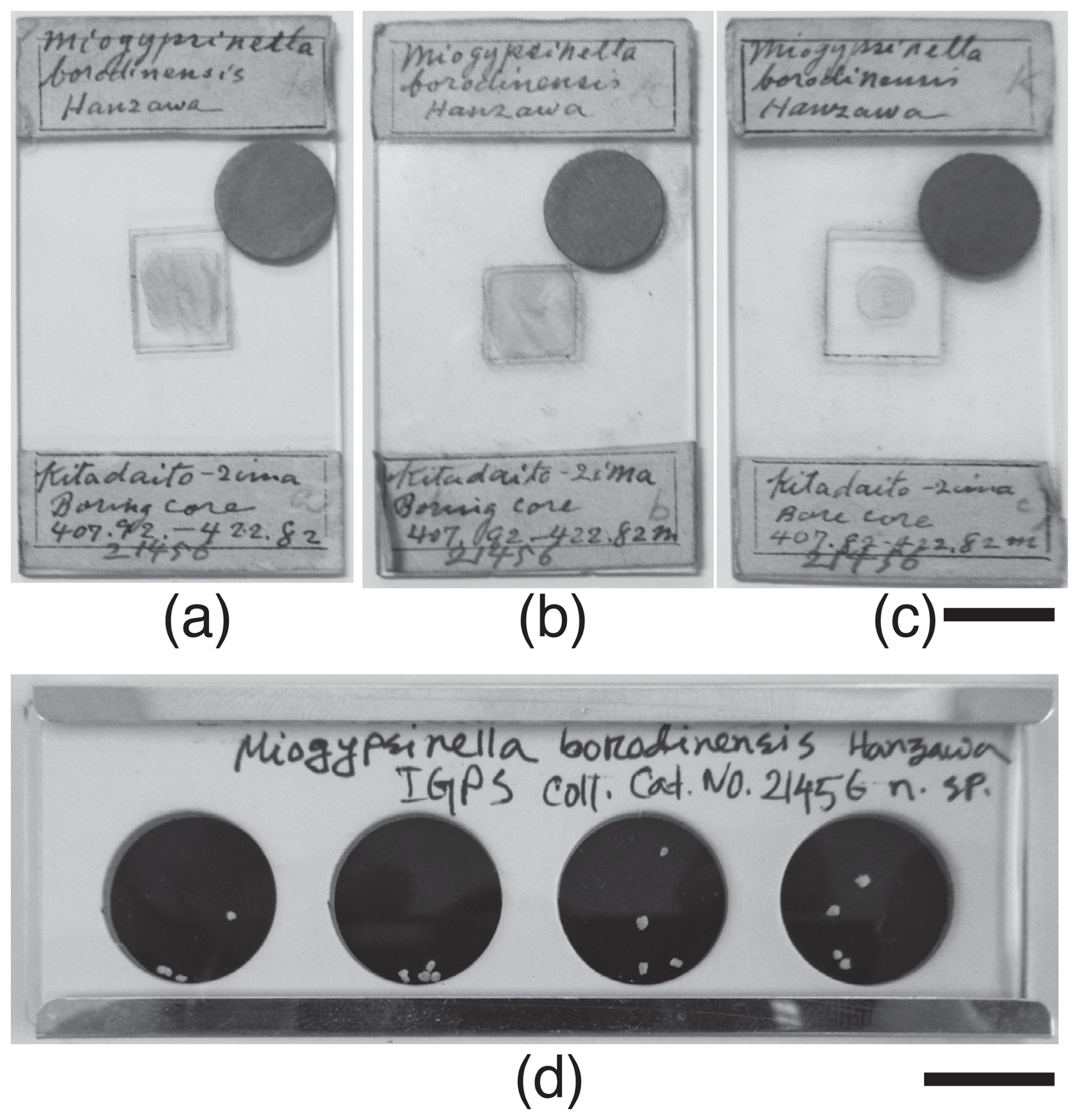
Figure 1Miogypsinella borodinensis Hanzawa, 1940, thin sections of types and isolated specimens; Hanzawa's collection, IGPS, Tohoku University, Sendai, Japan. (a) “Miogypsinella borodinensis Hanzawa, Kutadaito Jima Boring core 407.92–422.82 m, a”; lectotype, designated herein. (b) “Miogypsinella borodinensis Hanzawa, Kutadaito Jima Boring core 407.92–422.82 m, b”; paralectotype, designated herein. (c) “Miogypsinella borodinensis Hanzawa, Kutadaito Jima Boring core 407.92–422.82 m, c”; paralectotypes, designated herein. (d) “Miogypsinella borodinensis n. sp. Hanzawa, IGPS Coll. Cat. No. 21456”; paralectotype, designated herein. Scale bar represents 1 cm.
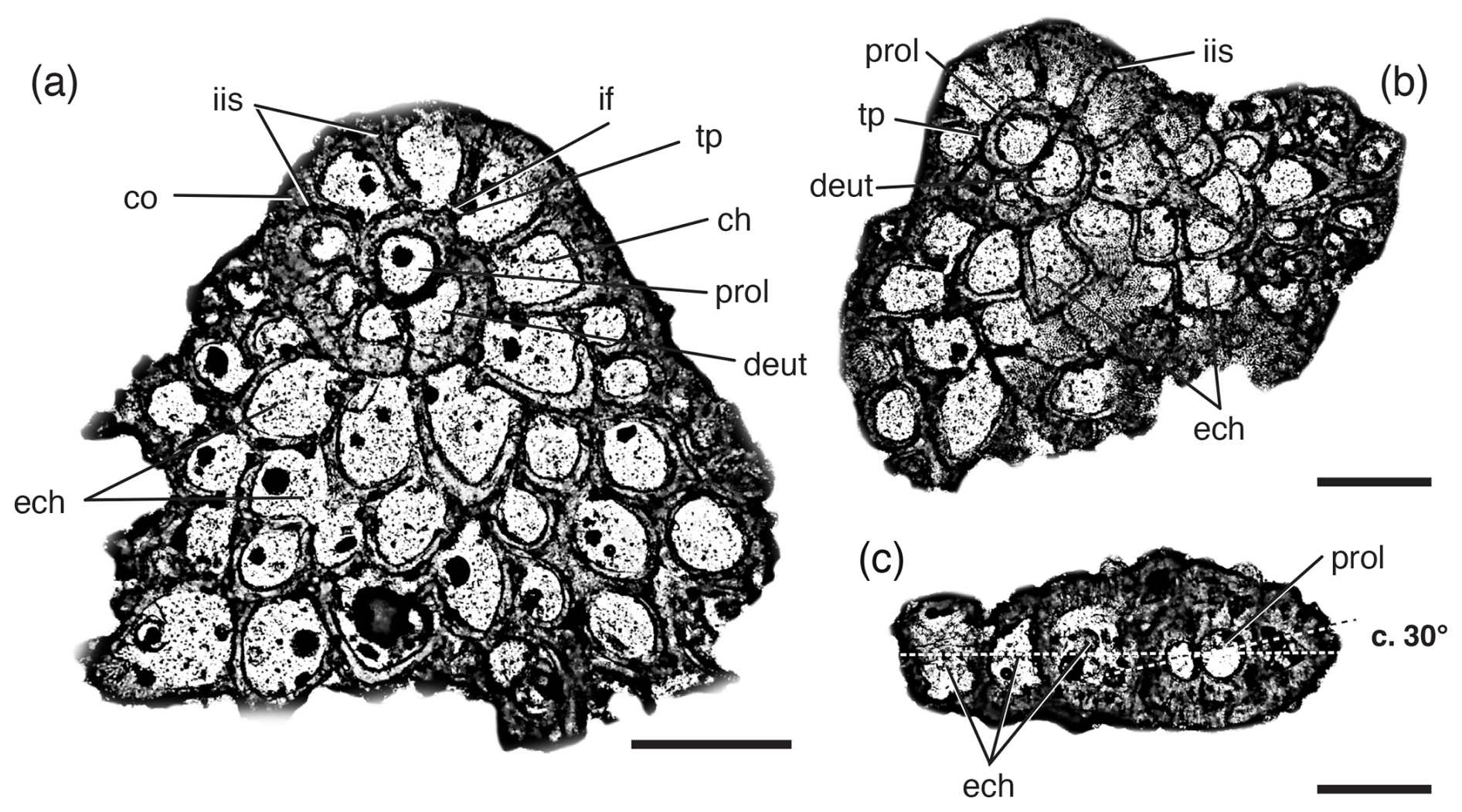
Figure 2Miogypsinella borodinensis Hanzawa, 1940; Hanzawa's collection; IGPS, Tohoku University, Sendai, Japan. (a) Equatorial section of an isolated specimen (Hanzawa, 1940, pl. 39, fig. 6). (b) Sub-equatorial section of an isolated specimen (Hanzawa, 1940, pl. 39, fig. 7). (c) Axial section of an isolated specimen (Hanzawa, 1940, pl. 39, fig. 8). Scale bar represents 0.250 mm. Abbreviations: ch, chamber; co, cover on intraseptal interlocular space; deut, deuteroconch; ech, equatorial chamberlet; if, intercameral foramen; iis, intraseptal interlocular space; prol, proloculus; tp, toothplate.
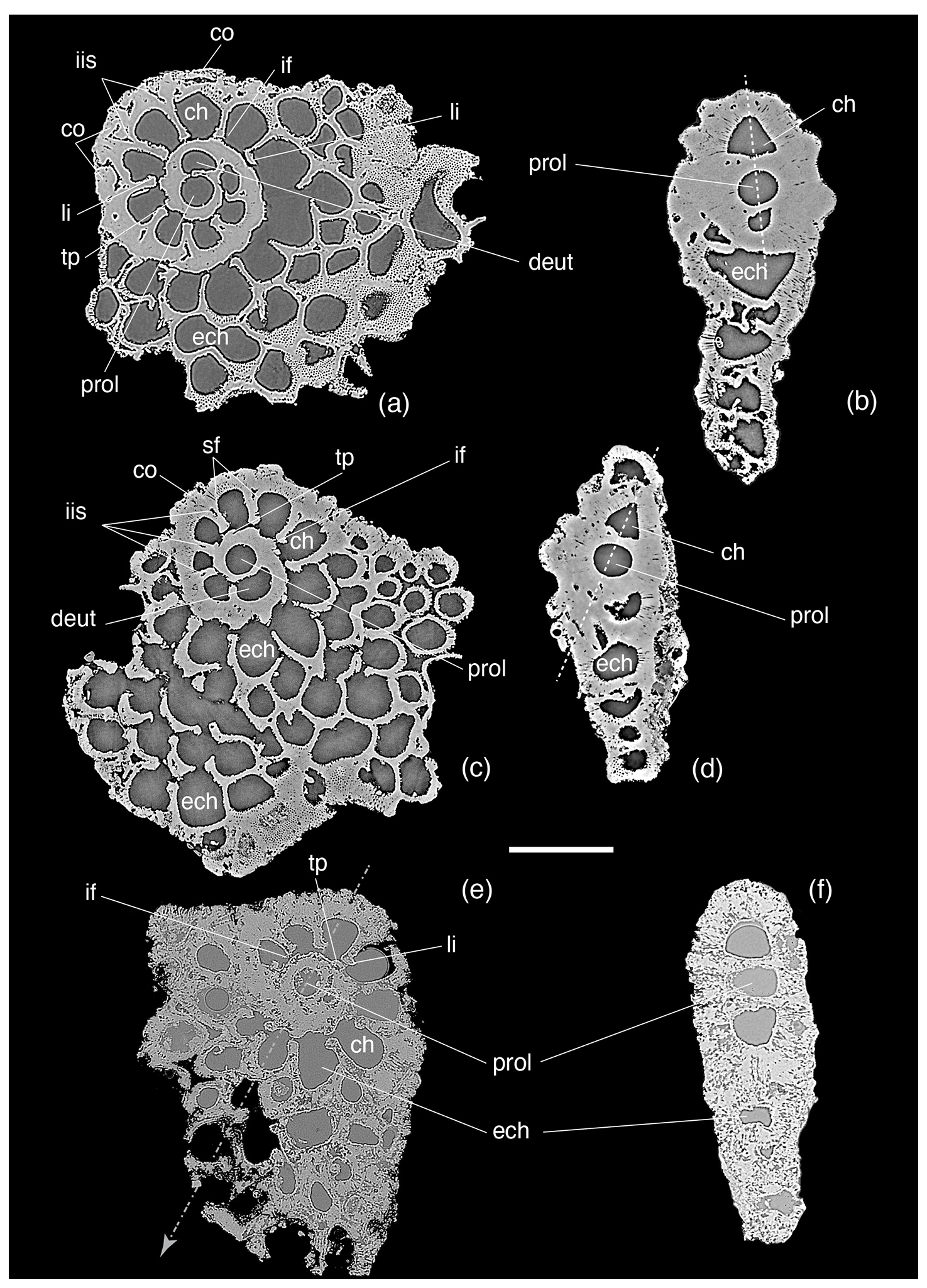
Figure 3Miogypsinella borodinensis Hanzawa, 1940; Hanzawa's collection; IGPS, Tohoku University, Sendai, Japan. Micro-computed tomographic analysis of megalospheric specimens. Scale bar represents 0.250 mm. For abbreviations see Fig. 2; li, apertural lip; sf, septal flap; tp, toothplate.
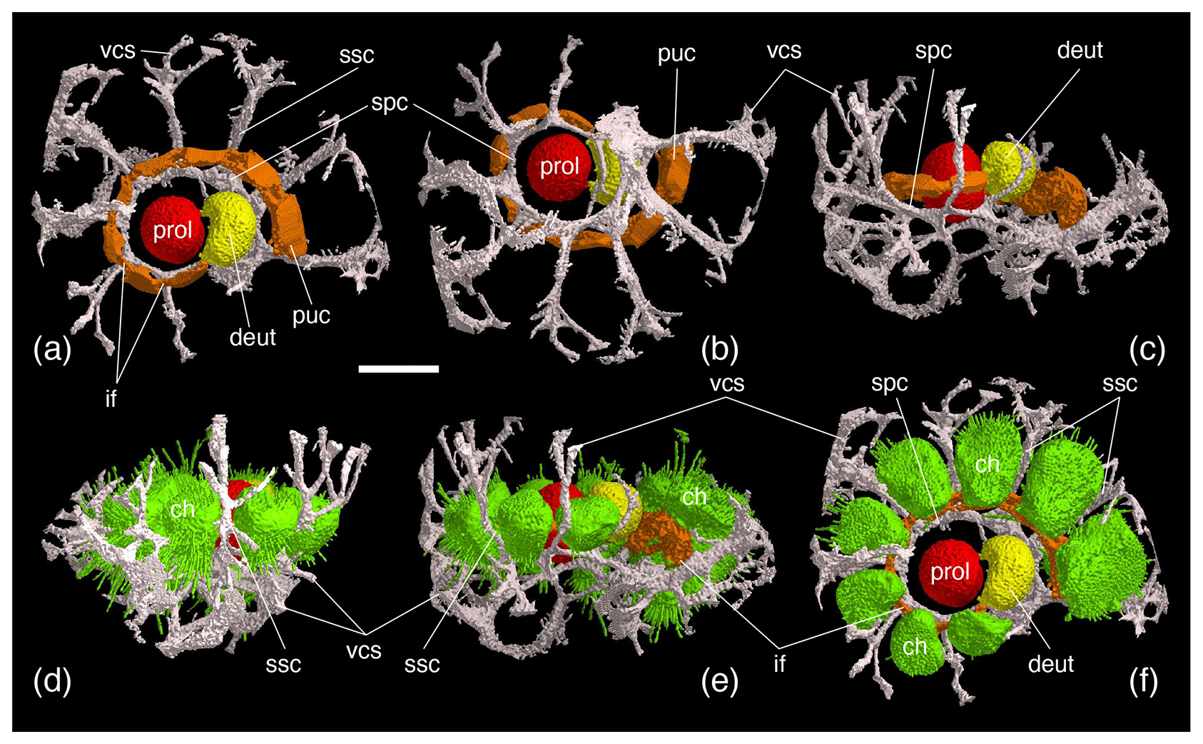
Figure 4Miogypsinella borodinensis Hanzawa, 1940; Hanzawa's collection; IGPS, Tohoku University, Sendai, Japan. Micro-computed tomographic scanning 3D-rendered models with shell removed (rendering by Shunichi Kinoshita). Primary spiral–umbilical canal is tubular (puc). The vertical canal system (vcs), issuing from the spiral canal (spc), connects successive volutions of the spiral canal. Subsutural canals (ssc; i.e. sutural fissures in de Bock, 1976) issue from deep interseptal spaces in the ultimate intercameral suture. For abbreviations see Fig. 2; sf, septal flap. Scale bar represents 0.100 mm.
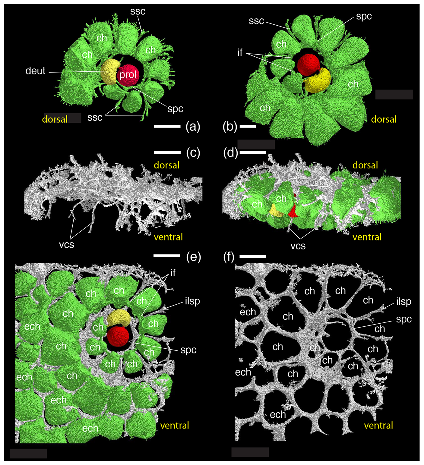
Figure 5Miogypsinella borodinensis Hanzawa, 1940; Hanzawa's collection; IGPS, Tohoku University, Sendai, Japan. Micro-computed tomographic scanning 3D-rendered models with shell removed (rendering by Shunichi Kinoshita). The ventral side of the shell is characterized by the vertical branches of the intraseptal canal system. Scale bars represent 0.100 mm. For abbreviations see Figs. 2–4.
Repository data. Thin sections labelled “Miogypsinella borodinensis Hanzawa, Kutadaito Jima Boring core 407.92–422.82 m, a”, “Miogypsinella borodinensis Hanzawa, Kutadaito Jima Boring core 407.92–422.82 m, b”, and “Miogypsinella borodinensis Hanzawa, Kutadaito Jima Boring core 407.92–422.82 m, c” (Fig. 1a) and isolated specimens in cell slide labelled “Miogypsinella borodinensis n. sp. Hanzawa, IGPS Coll. Cat. No. 21456” (Fig. 1b); housed at the IGPS, Tohoku University, Sendai, Japan.
Diagnosis. Nepionic stage with 7–12 chambers in trochospiral whorl near the shell apex. Proloculus ranges from 80 to 110 µm in diameter (Table 2). Angle γ ca. 140–165°. The nepionic planispiral whorl is inclined about ca. 30° with respect to the subsequent equatorial chambers.
Lectotype. Hanzawa (1940) did not designate a type. The illustrated syntypes of Hanzawa (1940) occur in three thin sections (Fig. 1a). Three isolated specimens are also present (Hanzawa, 1940; pl. 39, figs. 1–4). In accordance with Art. 74 of ICZN (1999), we hereby designate as lectotype the specimen in thin section “Miogypsinella borodinensis Hanzawa, Kutadaito Jima Boring core 407.92–422.82 m, a” (Fig. 1a), originally illustrated by Hanzawa (1940, pl. 39, figs. 5–6), with the purpose of clarifying the application of this name. The specimens of the other two thin sections illustrated in Fig. 1a thus become paralectotypes, as well as those in cell slide IGPS Coll. No. 21456.
Remarks. Hanzawa (1940) studied upper Oligocene material (see Iryu et al., 2010, for dated core data) collected from cores in the Kitadaito Jima borehole. The specimens illustrated in pl. 39, figs. 5–7, by Hanzawa (1940) show an apertural lip and subsutural canals, which are characters diagnostic for Miogypsinella. The micro-CT-scanned specimens show the occurrence of the cover on intraseptal interlocular space, intraseptal interlocular space, apertural lip, subsutural canals, septal flap, primary spiral–umbilical canal, thickness of the lateral walls, toothplate, and vertical canal system. In the analysed Hanzawa specimens, the proloculus diameter, angle γ, and inclination of the nepionic planispiral whorl confirm its status as a distinct species (Figs. 1–5, Table 2).
Hanzawa (1940, pp. 766–767, fig. 1) considered “Miogypsina complanata Schlumberger, 1900” described by Nuttall (1933) and by Barker and Grimsdale (1937) to be Miogypsinella based on the occurrence of sub-quadrate chambers in the spiral whorl covered by a compact layer of laminated structure on both sides of the shell (pp. 771–772). However, these characters cannot be considered valid for including M. complanata in Miogypsinella (e.g. Loeblich and Tappan, 1987; BouDagher-Fadel, 2018; Table 1).
Hanzawa (1940, p. 775) referred to “Miogypsina complanata Schlumberger” of Nuttall (1933, pl. 24, figs. 9, 11, 13–14) and to Miogypsinoides complanata of Barker and Grimsdale (1937; pl. 8, fig. 6), both from the upper Oligocene of eastern Mexico (Méson Formation; Vaughan and Cole, 1936; Wilson, 1987), to establish the new species Miogypsinella sanjosensis Hanzawa (p. 766). Hanzawa (1940) did not designate a holotype.
Later, Hanzawa (1957, p. 92) corrected the status of his species Miogypsinella sanjosensis, considering it to be a synonym of Miogypsinoides complanatus based on its “rotaloid stage”, which is not a distinctive character at the genus level (Table 1). The specimens of Nuttall (1933) show a biconvex test, 14 nepionic chambers (X=14), angle γ ca. 180°, and lateral chamberlets (pl. 24, fig. 11). The occurrence of lateral chamberlets rules out the ascription of these specimens to Miogypsinella.
In contrast, the specimens of Barker and Grimsdale (1937, pl. 8, fig. 6) show a spiral canal “… from which spring offshoots passing into, and along, the radial septa” (p. 163; i.e. the intraseptal interlocular space and subsutural canals). According to these shell characters, along with the septal flap, toothplate, and vertical canal system on ventral side only, those specimens can be ascribed to Miogypsinella rather than to Miogypsinoides (Table 1). No information about the collection storage was provided by Barker and Grimsdale (1937), and the original material described and illustrated could not be located. The studied isolated specimens were illustrated as equatorial (pl. 6, figs. 1–6; pl. 7, fig. 1) and axial sections (pl. 8, fig. 6). The nepionic stage shows 8–12 chambers in trochospiral whorl with an angle γ of ca. 130°. The proloculus ranges from 80 to 110 µm in diameter. The nepionic planispiral whorl direction angle is nearly 30° with respect to the later equatorial chambers. These characters correspond to Miogypsinella borodinensis (Table 2). The specimens illustrated by Barker and Grimsdale (1937) and the single drawing reported by Hanzawa (1940, fig. 3) cannot be morphologically separated from the types of Miogypsinella borodinensis, and, therefore, M. sanjosensis cannot be considered a distinct species.
By studying material from the Aquitanian of Bikini, Cole (1954) considered Miogypsinella borodinensis to be Miogypsinoides (p. 600) with no detailed systematic remarks (see also Cole, 1969, table 2). The illustrated specimens show more than 12 chambers in the nepionic stage and a proloculus diameter of ca. 80 µm. These characters are comparable to those of Miogypsinella ubaghsi rather than to those of M. borodinensis (Table 2).
Cole (1967, pl. 9, figs. 1, 3, 5, 8) reported as Miogypsinoides complanatus four specimens from the Chattian Méson Formation of eastern Mexico, from where the specimens of Barker and Grimsdale (1937) originated (see remarks above). In the equatorial sections of the illustrated specimens, the occurrence of ca. 10 nepionic chambers close to the shell apex, the intraseptal interlocular space, and subsutural canals suggests their ascription to Miogypsinella. This confirms that the specimens of Barker and Grimsdale (1937) and Cole (1967) from eastern Mexico represent the same species (i.e. Miogypsinella borodinensis).
Matsumaru et al. (2010) and Matsumaru (2017, p. 165) regarded Miogypsinella sanjosensis Hanzawa (1940) as a junior synonym of Miogypsinoides complanatus based on upper Oligocene materials from Turkey and the Philippine archipelago. The illustrated specimens clearly show heavily thickened lateral walls and vertical canal systems to both sides of the test confirming the ascription of these specimens to Miogypsinoides and ruling out a possible affinity to Miogypsinella.
Cole (1957c) described Miogypsinoides bantamensis from the Aquitanian of Saipan and considered Miogypsinella borodinensis to be its synonym (p. 328). The illustrated specimens (pl. 110, figs. 8–18; pl. 111, figs. 1–4) show heavily thickened lateral walls and vertical canal systems on both sides of the test, suggesting that this synonymy cannot be accepted.
Miogypsina (Miogypsinoides) cf. bermudezi recorded from the Chattian of Kutch (India; Raju, 1974) was illustrated by drawings (pl. 1, figs. 1–5); by their outer surfaces (pl. 3, figs. 1–2); and by three equatorial sections, which show nepionic chambers comparable in number (X=11–12; Raju, 1974; pl. 5, figs. 1–3) and in angle γ (130–150°) to those of M. borodinesis. This record of bermudezi likely corresponds to borodinensis (Tables 2 and 3). However, these Chattian records need further confirmation since no axial sections were illustrated (Raju, 1974). Random sub-axial sections of these Indian specimens show a thick lateral wall, suggesting possible affinities with Miogypsinoides.
Gedik (2018) reported Miogypsinella borodinensis from the Chattian of Malatya (Turkey). Gedik (2018) concluded that Miogypsinoides bantamensis, Miogypsinella borodinensis, and Miogypsinella bermudezi are synonyms of Miogypsinella borodinensis because of their number of nepionic chambers. This synonymy cannot be accepted due to the different shell characteristics of these species (Table 2). The thickness of the outer shell walls of the illustrated Malatya specimens suggests they are Miogypsinoides.
The Chattian records of Miogypsinella borodinensis from the western Tethys (Turkey; Sirel, 2003, 2015; Sirel and Gedik, 2011; Sirel and Işık, 2011; Hakyemez et al., 2016; Gedik, 2018) and the central Indo-Pacific (Borneo; BouDagher-Fadel, 2018) show planispiral nepionic chambers, heavily thickened lateral walls, and a vertical canal system to both sides of the test (Table 1). Therefore, these records belong to Miogypsinoides. The comparison between the proloculus diameter and the number of nepionic chambers of the recorded Miogypsinoides species in the Mediterranean areas (Schiavinotto and Benedetti, 2021, fig. 6) and M. borodinensis confirm this conclusion.
The single record of Miogypsinella borodinensis from the Chattian of Aquitaine (France; Cahuzac, 1984, pl. 1, fig. 5) does not clearly show its shell characters. Similarly, the single record of Miogypsinella from the Chattian of Brazil needs further confirmation since a thickened outer shell wall is likely present in the illustrated specimen (Alvarado Sierra et al., 2023).
Two drawings of axial sections of Miogypsinoides complanatus from the Oligocene of north-western Italy (as Miogypsina (Miogypsinoides) complanata in Ferrero Mortara (1987, fig. 4a–b) are similar to Miogypsinella borodinensis. However, due to the lack of information about the cover on intraseptal interlocular space, apertural lip, subsutural canals, and spiral canal, these specimens cannot be ascribed with certainty to Miogypsinella.
Geographical and stratigraphical distribution. Miogypsinella borodinensis appears in the Chattian in the central Indo-Pacific area (Figs. 6 and 7, Table 3). Central Indo-Pacific records of this species are from NE Borneo (BouDagher-Fadel et al., 2000a, b; BouDagher-Fadel and Price, 2013; BouDagher-Fadel, 2018) and Kitadaito Jima (Hanzawa, 1940, 1965), Saipan (Hanzawa, 1957), and the Bikini Atoll (Cole, 1954). The Central American records are from the Chattian of eastern Mexico (Barker and Grimsdale, 1937; Cole, 1967), Antigua, and Jamaica (Robinson and Persad, 1989; Robinson, 2004; Robinson et al., 2017; Mitchell et al., 2024). The last occurrences are from the Aquitanian of Kitadaito Jima (Hanzawa, 1965) and Bikini (Cole, 1954) (Fig. 6).
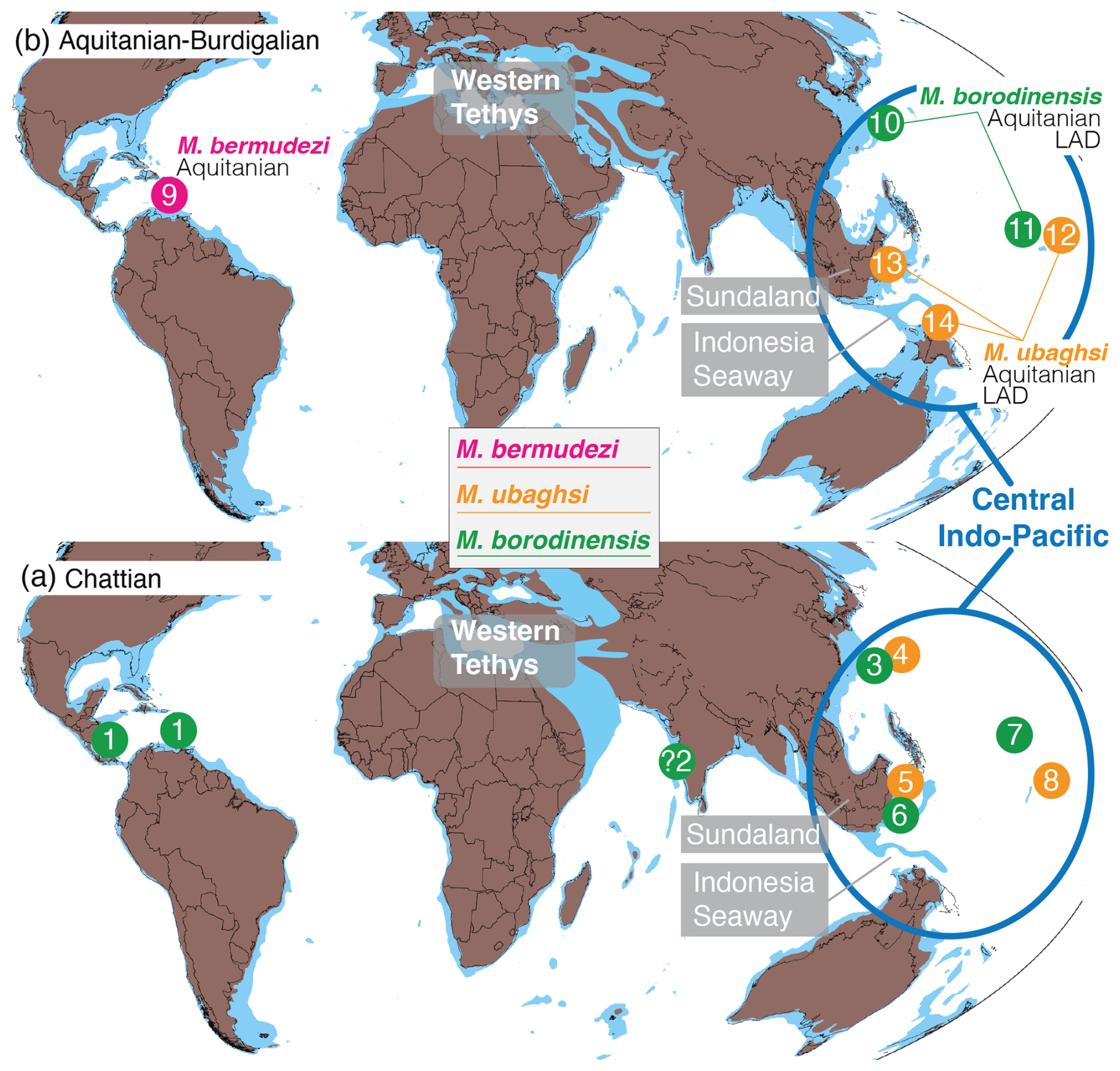
Figure 6Palaeogeographical locations of the early Oligocene (a)–early Miocene (b) Miogypsinella species. Abbreviations: Mbe, Miogypsinella bermudezi; Mbo, Miogypsinella borodinensis; Mu, Miogypsinella ubagshi; LAD, last appearance datum. In America, Miogypsinella borodinensis has so far been identified in the Chattian of eastern Mexico, Jamaica, and Antigua only (1, Barker and Grimsdale, 1937; Cole, 1967; Robinson, 2004; Robinson et al., 2017). Miogypsinella borodinensis appeared in the Chattian of Kitadaito Jima (3, Hanzawa, 1940), Saipan (7, Hanzawa, 1957), and north-eastern Borneo (6, BouDagher-Fadel et al., 2000a; BouDagher-Fadel and Price, 2013; BouDagher-Fadel, 2018). Its last occurrences are from the Aquitanian of Kitadaito Jima (10, Hanzawa, 1965) and Bikini (11, Cole, 1954). Miogypsinella ubaghsi ranges from the Chattian to the Aquitanian. This species occurs in the Chattian of Chichi Jima (4), north-eastern Borneo (5), and Eniwetok (8) (Tan, 1936a, b; Mohler, 1949; Cole, 1954, 1957b; Matsumaru, 2012; BouDagher-Fadel and Price, 2013; Mohiuddin et al., 2000; BouDagher-Fadel, 2018) as well as the Aquitanian of Bikini (12), north-eastern Borneo (13), and Papua New Guinea (14) (Tan, 1936a; Cole, 1954; Hanzawa, 1940; BouDagher-Fadel et al., 2000a; BouDagher-Fadel and Price, 2013). The single record of Miogypsinella bermudezi is from the early Miocene of Cuba (9, Drooger, 1951). Numbers refer to localities. 1: Barker and Grimsdale (1937; Mbo, eastern Mexico), Cole (1938, 1957a, 1967; Mbo, eastern Mexico, Panama). 2: Raju (1974; Mbo, Kutch). 3: Hanzawa (1940; Mbo, Kitadaito Jima), Matsumaru (2012; Mbo, Kitadaito Jima), this study. 4: Matsumaru (2012; Mbo, Mu, Chichi Jima). 5: BouDagher-Fadel et al. (2000a, b; Mbo, Mu, NE Borneo), BouDagher-Fadel and Price (2013; Mbo, Borneo), BouDagher-Fadel (2018; Mbo, NE Borneo). 7: Hanzawa (1957; Mbo, Saipan). 8: Hanzawa (1962; Mu, Eniwetok). 9: Drooger (1951; Mbe, Pinar del Rio, Cuba). 10: Hanzawa (1965; Mbo, Kitadaito Jima). 11: Cole (1954; Mbo, Bikini). 12: Cole (1954; Mu, Bikini). 14: BouDagher-Fadel et al. (2000a; Mu, NE Borneo, Papua New Guinea), BouDagher-Fadel and Price (2013; Mu, NE Borneo, Papua New Guinea), BouDagher-Fadel (2018; Mu, Papua New Guinea). Palaeogeographical maps modified from Hall (2013) and Kocsis and Scotese (2021). Flooded continental areas are indicated with light blue; terrestrial areas are coloured brown.
-
Miogypsinella bermudezi (Drooger, 1951)
-
1951 Miogypsina (Miogypsinella) bermudezi n. sp., Drooger, pp. 357–359, figs. 1a–6.
-
1952 Miogypsina bermudezi Drooger; Drooger, p. 47.
-
? 1957 Miogypsina (Miogypsinoides) bermudezi Drooger; Akers and Drooger, pp. 670, 674.
-
1957 M. bermudezi (Drooger) (1951) (= M. ubaghsi Tan); Hanzawa, p. 86.
-
1993 Miogypsina bermudezi; Drooger, fig. 43 (left), fig. 44 (left).
-
non 2003 Miogypsinella bermudezi Drooger; Sirel, pl. 14, figs. 1–27.
Type reference and figures. Miogypsina (Miogypsinella) bermudezi Drooger, 1951, pp. 357–359, figs. 1a–6; figs. 1a–c, 2a–c, 3a–b (holotype).
Repository data. Unfortunately, the original material described by Drooger (1951) could not be located at the Department of Earth Sciences, Faculty of Geosciences, Utrecht University.
Diagnosis. Nepionic stage with 13–19 chambers in trochospiral whorl near the shell apex. Proloculus ranges from 50 to 70 µm in diameter (Table 2). Angle γ ca. 300°. The nepionic planispiral whorl is nearly horizontal with respect to the subsequent equatorial chambers.
Remarks. Drooger (1951) introduced this Miogypsinella species from the Miocene of Cuba. The holotype is an isolated specimen illustrated by drawings (figs. 1a–c, 2a–c, 3a–b), whereas three specimens (paratypes) are represented by equatorial (fig. 4) and axial sections (figs. 5–6). The paratypes show intraseptal interlocular space, subsutural canals, septal flap, primary spiral canal, subsutural canals, thickness of the lateral walls, toothplate, and a vertical canal system, indicating that they belong to Miogypsinella. They show 12 nepionic chambers, arranged in a planispiral whorl nearly horizontal (i.e. planispiral whorl direction degree 0°; figs. 5–6), with a proloculus 50–70 µm in diameter (Drooger, 1951, p. 359). The angle γ is ca. 300° (Drooger, 1951, fig. 4).
This species suffered the taxonomic uncertainties regarding the distinction between Miogypsinoides and Miogypsina. Drooger (1952, 1993) ascribed M. bermudezi to Miogypsina but afterwards regarded the species as Miogypsinoides bermudezi (1963, fig. 4; Drooger and Raju, 1973). The distinctive short nepionic stage and the “difference in thickness of the solid side wall” with respect to Miogypsinoides (Drooger, 1993, p. 76, 89, fig. 43) suggest a separate status of this species as Miogypsinella bermudezi. This status is further confirmed by the occasional occurrence of intraseptal interlocular space and subsutural canals, referred to by Drooger (1993, p. 83) as “lateral chambers”.
Miogypsinella bermudezi differs from M. borodinensis in having a smaller proloculus (50–70 µm versus 90 µm), a higher number of nepionic chambers (X=13–19 versus X=7–12), a higher angle γ (ca. 300° versus 140–165°), and a horizontal early trochospiral whorl (Table 2). Miogypsinella bermudezi is similar to M. ubaghsi (Table 2) except for the proloculus diameter (50–70 µm versus 80–130 µm).
Since no illustrations were published, Early Miocene records from the Louisiana Gulf Coast (Akers and Drooger, 1957; see Poag, 1975, for stratigraphy) and Puerto Rico (Gordon, 1961) need further study. Nonetheless, Akers and Drooger (1957) described “thicker side walls and pustules” which do not support the ascription to the thinner-walled Miogypsinella.
Chattian specimens from Antigua and Jamaica (Robinson, 2004) are characterized by thinner lateral side walls, proloculus size, and number of nepionic chambers comparable to those of Miogypsinella borodinensis rather than to M. bermudezi (Table 2).
Miogypsinoides nigeriana (Küpper, 1960), from the Burdigalian of Cameroon, is characterized by very thin lateral walls and a sub-horizontal planispiral nepionic whorl resembling those of Miogypsinella bermudezi. However, because in Miogypsinoides nigeriana the canal system occurs at both sides of the test, this species was correctly ascribed to Miogypsinoides (Drooger, 1966). The Chattian record of Miogypsinella bermudezi from the western Tethys (Turkey; Sirel, 2003, pl. 14, figs. 1–27) shows shell characters distinctive of Miogypsinoides (Table 1).
Stratigraphical distribution. Drooger (1951) attributed his material from Baños in Pinar del Rio Province, western Cuba, to the “Early Middle Oligocene” (p. 359). According to the stratigraphical reassessment of the Oligocene–Miocene stratigraphy of Cuba, the Baños succession was formalized as the Baños Formation (Iturralde-Vinent, 1972). This formation consists of two members: the lower member (Oligocene–lowermost Miocene) with deep-water benthic foraminifera and lepidocyclinids and the upper member (Miocene) consisting of sandy limestone with lepidocyclinids, nummulitids, and miogypsinids. The material of Drooger (1951) probably came from the upper member, early Miocene in age (Figs. 6–7, Table 3).
-
Miogypsinella ubaghsi (Tan, 1936)
-
1936a Miogypsinoides ubaghsi n. sp. Tan, pp. 47–48, pl. 1, figs. 1, 7.
-
non 1936a Miogypsinoides ubaghsi n. sp. Tan, pl. 1, figs. 3–6.
-
1940 Miogypsinella ubaghsi (Tan); Hanzawa, pp. 767–768, 775, text-fig. 4.
-
1949 Miogypsina (Moides) ubaghsi; Mohler, p. 526.
-
1954 Miogypsinoides ubaghsi Tan; Cole, pp. 603–604, pl. 221, ? fig. 5, figs. 9–17, no fig. 18, pl. 222, figs. 13, 15, no fig. 14.
-
1954 Miogypsinoides borodinensis (Hanzawa); Cole, p. 600, pl. 221, figs. 6–8.
-
1957b Miogypsinoides ubaghsi Tan; Cole, pp. 770–771, pl. 243, figs. 5–16.
-
non 1957b Miogypsinoides ubaghsi Tan; Cole, pl. 243, figs. 10–11, 13–14, 17–19.
-
1957 Miogypsinella bermudezi (Drooger); Hanzawa, p. 86.
-
? 1962 Miogypsinoides ubaghsi Tan; Hanzawa, pl. 7, figs. 16–17, 19 (drawings).
-
? 1984 Miogypsinoides ubaghsi Tan Sin Hok; Cahuzac, pl. 1, fig. 2.
-
? 1986 Miogypsinoides ubaghsi; Premoli Silva, pl. 1, figs. 1–3, 9.
-
? 1981 Miogypsinoides ubaghsi; Premoli Silva and Brusa, pl. 12, figs. 12–13, pl. 13, fig. 5.
-
? 1991 Miogypsinoides ubaghsi; Gibson and Margerum, fig. 8.
-
1996 Miogypsinella boninensis n. sp.; Matsumaru, pp. 50, 52, 54, pl. 7, figs. 12–13; figs. 23–24.
-
non 1996 Miogypsinella boninensis n. sp.; Matsumaru, pl. 5, figs. 1–7; pl. 6, figs. 1–12; pl. 7, figs. 1–11, 14–16.
-
2000a Miogypsinella ubaghsi (Tan Sin Hok); BouDagher-Fadel et al., pl. 3, figs. 1–2.
-
2000a Miogypsinella boninensis Matsumaru; BouDagher-Fadel et al., pl. 2, figs. 1–2, 4.
-
2000 Miogypsinella ubaghsi (Tan, 1936; Mohiuddin et al., p. 199, figs. 7.2, 8.2, 8.3.
-
2005 Miogypsinella boninensis Matsumaru, 1996; Sharaf et al., p. 10, pl. 1, fig. 1.
-
2018 Miogypsinella ubaghsi (Tan Sin Hok); BouDagher-Fadel, pl. 7.14, figs. 4, 8.
-
non 2020 Miogypsinella ubaghsi; Teillet et al., fig. 5A.
Type reference and figures. Tan (1936a), pp. 47–48, pl. 1, figs. 1–3, 7.
Repository data. The original material described and illustrated by Tan (1936a) could not be located.
Diagnosis. Nepionic stage with 13–16 (maximum 24) chambers in a trochospiral whorl near the shell apex. Proloculus ranges from 80 to 130 µm in diameter (Table 2). Angle γ ca. 330° (maximum 400°). The nepionic planispiral whorl is nearly horizontal with respect to the equatorial chambers.
Remarks. Tan (1936a) established the new species Miogypsinoides ubaghsi from the Aquitanian of Indonesia. Hanzawa (1940) considered M. ubaghsi to be congeneric with Miogypsinella borodinensis, both taxa having “a juvenarium of rotaloid spire and umbilical plugs” (p. 767). The occurrence of subsutural canals and a vertical canal system only in the ventral shell side in the types of ubaghsi (Tan, 1936a, pl. 1, figs. 1–3, 7) indicates that it belongs to Miogypsinella. Miogypsinella ubaghsi differs from the other species in having a larger proloculus, the highest number of nepionic chambers, and the lowest angle γ (Table 2).
In the Aquitanian of Eniwetok, Cole (1957b) identified Miogypsinoides ubaghsi with no detailed systematic remarks (see also Cole, 1969, table 2). The illustrated specimens show a thickened lateral shell wall, indicating their ascription to Miogypsinoides.
Miogypsinella boninensis Matsumaru, 1996, described from the Chattian of Minami-jima (Japan), was considered a species separated from M. ubaghsi essentially by having a proloculus ca. 90–140 µm in diameter and 23–28 nepionic chambers (Matsumaru, 1996, p. 52). However, the proloculus diameter and the number of the nepionic chambers, along with an unclear angle γ (Matsumaru, 1996, pl. 5, figs. 1–7; pl. 6 fig. 6, holotype; fig. 23/4), question the separation of the two species (see also Matsumaru, 1996, p. 54, no illustrated axial sections) and suggest that M. boninensis likely belongs to Miogypsinoides.
Cahuzac (1984) illustrated a single specimen from the Chattian of Aquitaine (France) but did not provide any information about the shell characters.
Most of the Miogypsinella ubaghsi (as Miogypsinoides) records are inadequately illustrated, and some shell characters do not fit with the species circumscription, as they show heavily thickened lateral walls and vertical canal systems to both sides of the test (Table 1; Cole, 1954, 1957b; Premoli Silva, 1986; Premoli Silva and Brusa, 1981; Gibson and Margerum, 1991; Teillet et al., 2020).
Stratigraphical distribution. The material of Tan (1936a) was collected from the Aquitanian of Indonesia. This species has been recorded from the Chattian in Borneo, Eniwetok, Chichi Jima, Komahashi-daini Seamount, Kitadaito Jima, and Kyushu-Palau ridge (Mohler, 1949; Hanzawa, 1962; Matsumaru, 1996, 2012; Mohiuddin et al., 2000) as well as the Aquitanian in Papua New Guinea (Cole, 1954, 1957b; BouDagher-Fadel et al., 2000a; BouDagher-Fadel and Price, 2013; BouDagher-Fadel, 2018) (Figs. 6 and 7, Table 3).
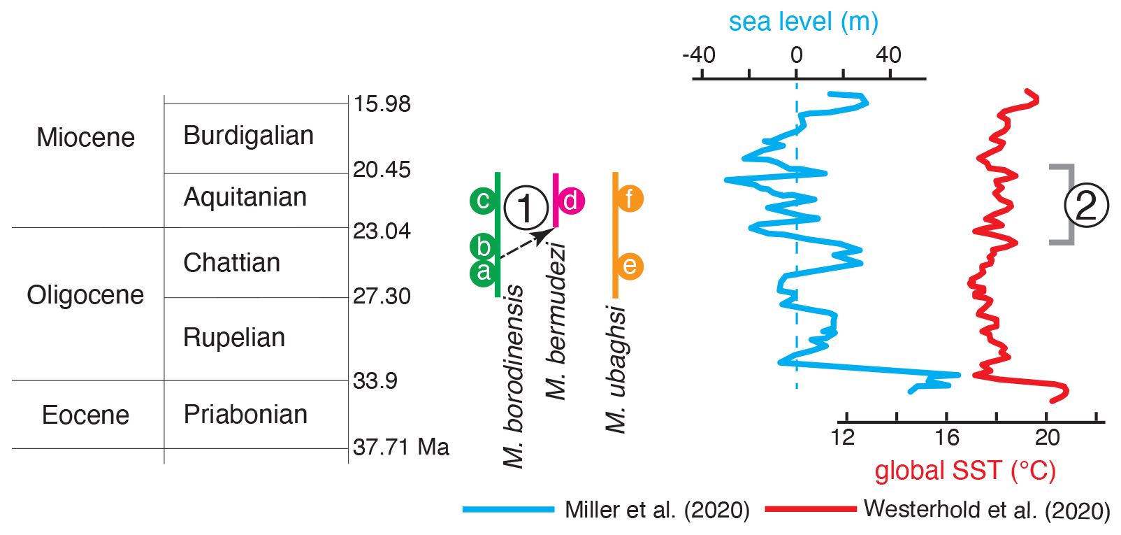
Figure 7Biostratigraphical patterns of the lower Oligocene–lower Miocene Miogypsinella species compared with the global eustatic curves and global sea surface temperature (SST) estimates. Miogypsinella borodinensis (a, b) and M. ubaghsi (e) appeared during the Chattian peaks in sea level curve (e.g. Miller et al., 2020) and global SST (e.g. Westerhold et al., 2020). The occurrence of Miogypsinella borodinensis in the Chattian of eastern Mexico (b) is interpreted as the result of Pacific migrants likely following an eastward path to Central America. Miogypsinella bermudezi (d) descended from M. borodinensis (1). Miogypsinella borodinensis (c) and M. ubaghsi (f) disappear near the Aquitanian–Burdigalian eustatic rise, which nearly coincides with the initiation of the Indonesian Seaway restriction and the abrupt deposition of deeper-marine clays in Southeast Asia (ca. 23–20 Ma; 2). See text and Table 3 for details; a: Cole (1954), BouDagher-Fadel (2018), BouDagher-Fadel et al. (2000a), BouDagher-Fadel and Price (2013), Hanzawa (1940, 1957), and Raju (1974); b: Barker and Grimsdale (1937), Cole (1967), Hanzawa (1940); c: Hanzawa (1965); d: Drooger (1951); e: Mohler (1949), Cole (1957b), Hanzawa (1962), Matsumaru (1996, 2012), BouDagher-Fadel et al. (2000a), Mohiuddin et al. (2000), Sharaf et al. (2005); f: BouDagher-Fadel (2018), BouDagher-Fadel and Price (2013), Cole (1954), Tan (1936a, b). Timescale after Cohen et al. (2025).
-
Genus Miogypsinoides Yabe and Hanzawa, 1928
Type species. Miogypsina dehaartii van der Vlerk, 1924, pp. 429–432, figs. 1a–c, figs. 2–3.
Diagnosis. Embryo near test apex; nepionic chambers in a single planispiral whorl with spherical proloculus and reniform protoconch; asymmetrical equatorial chamberlets; septal flap, intraseptal interlocular space with cover, vertical canals extended to both sides of the shell; distinctively lamellar, thick lateral walls.
-
Miogypsinoides lateralis Hanzawa, 1940
Figures 8 and 9. -
1940 Miogypsinoides lateralis n. sp. Hanzawa, p. 783, pl. 39, figs. 10–14.
-
1953 Miogypsinoides formosensis Yabe and Hanzawa; Cole et al., pl. 14, fig. 8.
-
1957 Miogypsinoides lateralis Hanzawa; Hanzawa, pp. 92–93.
-
1957 Miogypsinoides dehaartii (Van der Vlerk); Cole, pl. 111, figs. 6, 11.
-
1957 Miogypsinoides bantamensis Tan; Cole, pl. 110, fig. 15.
-
1962 Miogypsinoides lateralis Hanzawa; Hanzawa, pl. 7, figs. 9, 13.
-
1969 Miogypsinoides dehaartii (van der Vlerk) (with “M. lateralis” kind of embryonic apparatus”); Cole, pl. 1, figs. 5–6, 9, 11–12, 20.
-
1974 Miogypsina (Miogypsinoides) bantamensis Tan Sin Hok; Adams and Belford, p. 496, pl. 73, figs. 8–11.
Type reference and figures. Miogypsinoides lateralis Hanzawa, 1940, p. 783, pl. 39, figs. 10–11 (isolated specimens), 12 (axial section), 13 (equatorial section), 14 (equatorial section, detail of the nepionic stage).
Repository data. Thin sections labelled “Miogypsinoides lateralis Hanzawa, Kutadaito Jima Boring core 363.76–371.83 m, a” (Fig. 8a) and isolated specimens in cell slide labelled “AQUITANIAN Kita-Daito-jima Deep well. 331.52–338.57 Deep Hypotype Miogypsinoides lateralis Hanzawa IGPS. Coll. Cat. No. 21491” and “Miogypsinoides lateralis Hanzawa n. sp. Hypotype 351.52–338.57 m IGPS Cat. 21491” (Fig. 8d); housed at the IGPS, Tohoku University, Sendai, Japan.
Diagnosis. Nepionic stage with seven to eight chambers in a planispiral whorl. Sub-spheroidal proloculus ca. 160 µm in diameter followed by reniform deuteroconch, ca. 175 µm wide (Fig. 9b, d and e). Arc length of nepionic spiral whorl starting from embryonic chambers and ending at the apical point of test of ca. 90°. Nepionic chambers are connected by a primary spiral canal (Fig. 9b, d and e). Intraseptal interlocular space formed between the posterior bilamellar wall of a chamber and the distal bilamellar wall of the preceding one as a result of a deeply sunken suture (Fig. 9c–e). Intraseptal space is open to the exterior along its margins either continuously or through openings between points of marginal adherence of consecutive lateral chamber walls (Fig. 9c–e). Toothplates develop from an intercameral foramen to an aperture and are attached to both (Fig. 9d). These toothplates are shaped to form a single fold with a free distal end and distally protruding into the aperture. Septal flaps are part of the inner lamella covering the preceding septal face. By its adherence to the septal face, the septal flap produces a trilamellar septum (Fig. 9d).
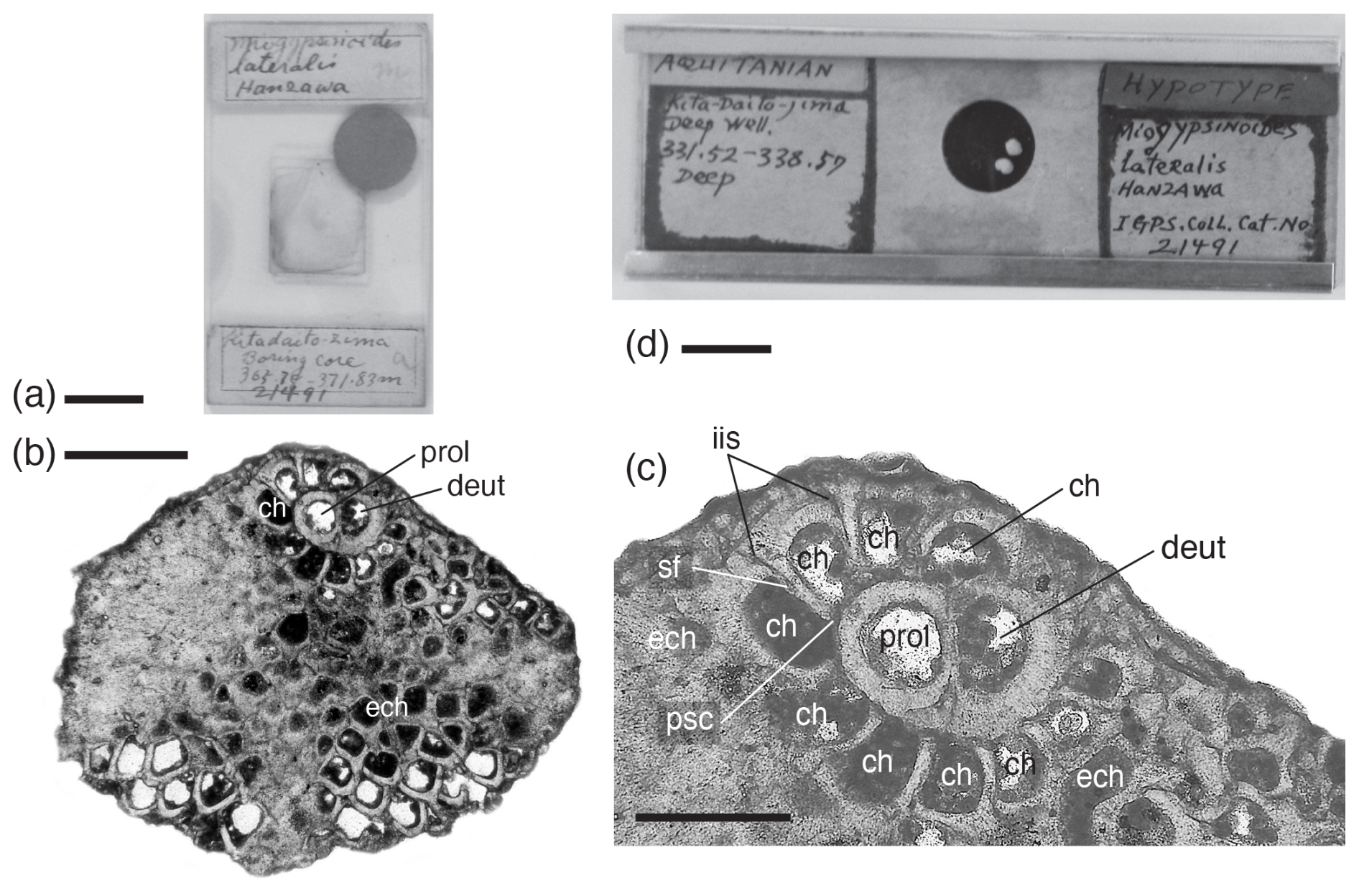
Figure 8Miogypsinoides lateralis Hanzawa, 1940, thin section of types (a–c) and isolated specimens (d); Hanzawa's collection; IGPS, Tohoku University, Sendai, Japan. (a) “Miogypsinoides lateralis Hanzawa, Kitadaito-zima Boring core 363.76–371.83 m, 21491”. (b, c) Equatorial section of an isolated specimen (Hanzawa, 1940, pl. 39, figs. 13–14. (d) “Miogypsinoides lateralis Hanzawa, IGPS. Coll. Cat. No. 21491, Kita-daito-jima, Deep well, 331.52–338.57, deep, Aquitanian”. Scale bar represents 1 cm in (a) and (d), 0.500 mm in (b), and 0.250 mm in (c). For abbreviations, see Fig. 2.
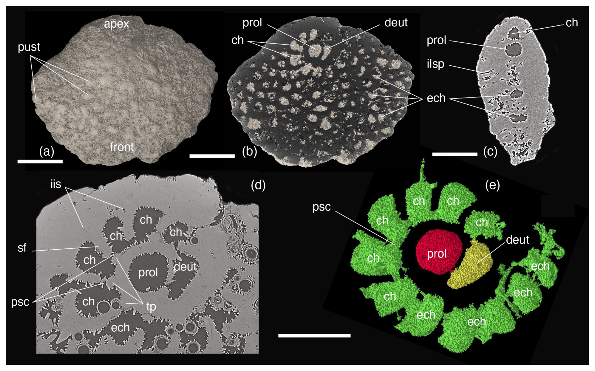
Figure 9Miogypsinoides lateralis Hanzawa, 1940; Hanzawa's collection; IGPS, Tohoku University, Sendai, Japan. Micro-computed tomographic analysis of megalospheric specimens showing outer shell morphology (a) and equatorial (b) and axial (c) sections. (d, e) Micro-computed tomographic scanning 3D-rendered models with shell removed (rendering by Shunichi Kinoshita). Scale bar represents 0.500 mm in (a)–(c) and 0.250 mm in (d) and (e). For abbreviations, see Fig. 2.
Lectotype. Hanzawa (1940) did not designate a type. The original material illustrated by Hanzawa (1940) occurs in one thin section (Fig. 8a). In accordance with Art. 74 of ICZN (1999), we hereby designate as lectotype the specimen in thin section “Miogypsinoides lateralis Hanzawa, Kutadaito Jima Boring core 363.76–371.83 m, a” (Fig. 8a–c), originally illustrated by Hanzawa (1940, pl. 39, figs. 13–14), with the purpose of clarifying the application of this name. The isolated specimens in cell slides thus become paralectotypes (Fig. 8d).
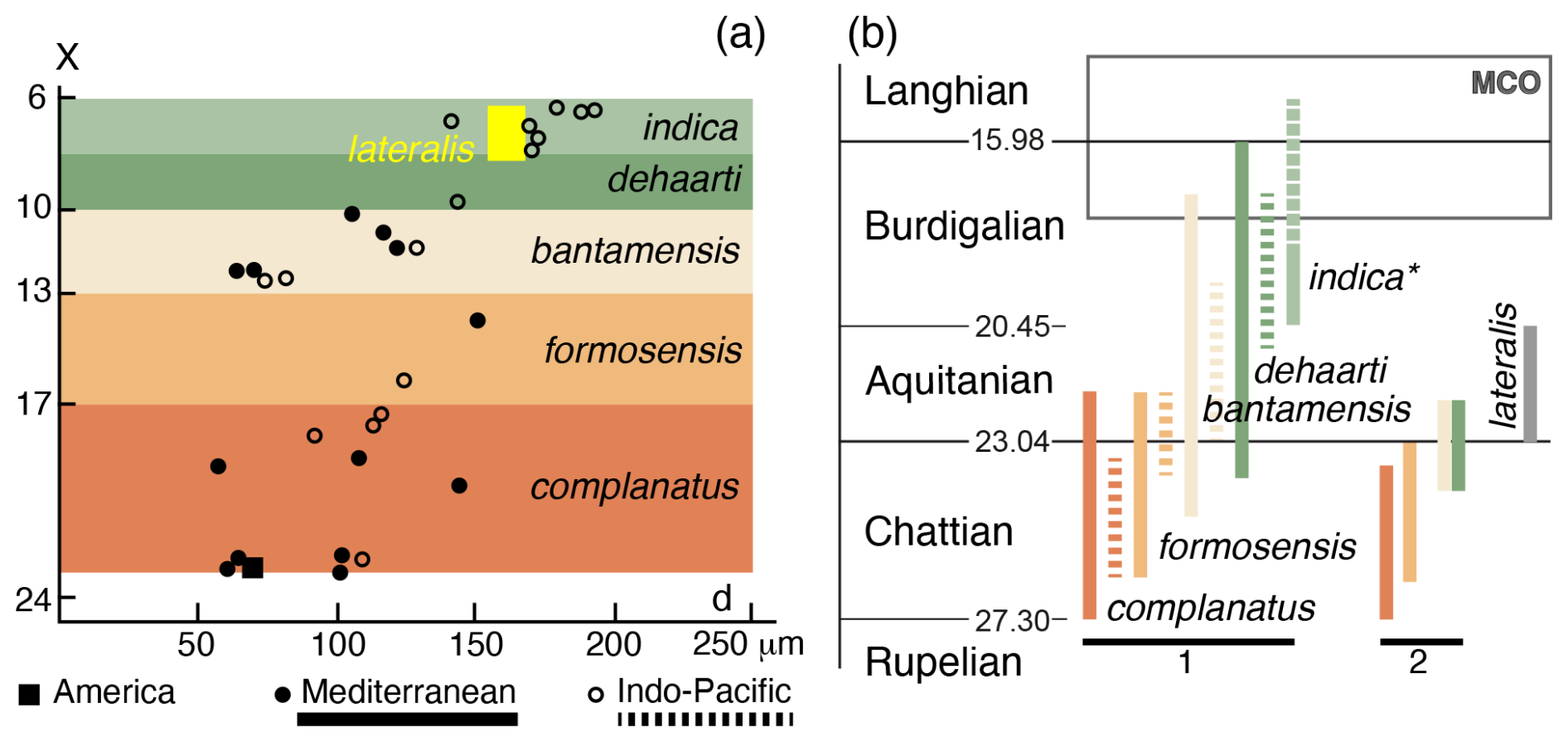
Figure 10(a) Plot of mean protoconch diameter (d) and number of nepionic chambers (X) for the Miogypsinoides species from American, Mediterranean, and Indo-Pacific areas (Drooger and Raju, 1973; Drooger, 1993). Although values of d−X of Miogypsinoides lateralis fall into the lower Burdigalian M. indica range, M. lateralis clearly differs from M. indica in having a larger angle γ (90° versus 17°). The Aquitanian Miogypsinoides lateralis likely represents an exception to the nepionic acceleration trend. (b) Stratigraphical distributions of Miogypsinoides species and location of M. lateralis from the Aquitanian of Kitadaito Jima. 1: Lunt and Allan (2004), BouDagher-Fadel and Price (2013), BouDagher-Fadel (2018). 2: Drooger and Laagland (1986), Cahuzac and Poignant (1987; as to bantamensis–dehaarti), Özcan et al. (2009). * Raju (1974, fig. 38) localized M. indica in the lower Burdigalian, whereas BouDagher-Fadel (2018, fig. 7.16) considered the species to be upper Burdigalian–lower Langhian in age. MCO, Miocene Climatic Optimum. Timescale after Cohen et al. (2025).
Remarks. The specimens illustrated in pl. 39, figs. 12–14 (Hanzawa, 1940) show the thickness of the lateral wall and the ventral canal system to either side of the shell, which are characters diagnostic for Miogypsinoides (Table 1). The occurrence of nepionic chambers in a single planispiral whorl, intraseptal interlocular space, thickened lateral walls with vertical and lateral canal systems to both sides, and equatorial ogival chambers confirms that the type specimens belong to the genus Miogypsinoides (de Bock, 1976; Loeblich and Tappan, 1987; Drooger, 1993; Table 1).
Although considered a junior synonym of Miogypsinoides bantamensis (Cole, 1957c, p. 339; Drooger, 1963, p. 346), M. lateralis differs from M. bantamensis in having fewer nepionic chambers (6–7 versus 11–14). Based on specimens from Saipan, Cole (1967) reconsidered Miogypsinoides lateralis to be a distinct species, with the “lateralis kind of specimens” occurring with the “bantamensis” and “dehaartii” “kinds”. Cole (1969) identified Miogypsinoides lateralis along with M. bantamensis, M. dehaartii, and M. mauretanicus in deep drill cores in the Midway Atoll. Despite stating that Miogypsinoides lateralis is easily distinguishable from M. bantamensis, Cole (1969, p. C11) proposed to group both species into M. dehaarti (p. C12) because they were found “constituting a continuous series” in one part of the studied region. This proposal is not supported by any morphologic characters, which clearly differentiate Miogypsinoides lateralis from the other species. Adams and Belford (1974) considered Miogypsinoides lateralis to be a synonym of M. bantamensis with no discussion on systematic remarks.
Based on Aquitanian specimens from Shikoku (Japan), Matsumaru et al. (1993) considered Miogypsinoides lateralis to be a synonym of Miogypsinoides dehaartii. However, the illustrated axial sections do not show specific diagnostic characters, and the ascription of specimens is uncertain. Later, Matsumaru et al. (2010, p. 454) and Matsumaru (2017, p. 166) also regarded Miogypsinoides lateralis as a synonym of M. bantamensis. The described and illustrated specimens clearly do not belong to Miogypsinoides lateralis as they show 10–14 nepionic chambers and an angle γ of ca. 150°. Raju (1974) described Miogypsinoides indica from the lower Burdigalian of Kutch and Saurastra in India. This species is comparable to Miogypsinoides lateralis in the number of nepionic chambers (five to eight versus seven to eight) and the protoconch diameter (ca. 193 µm versus 175 µm) (Fig. 10a). However, the angle γ (17° versus 90°) clearly separates the two species.
Stratigraphical distribution. The genus Miogypsinoides ranges from the Rupelian to the Chattian in America (Drooger, 1993; BouDagher-Fadel and Price, 2013; BouDagher-Fadel, 2018), from the Chattian to the Burdigalian in the Mediterranean (Drooger and Laagland, 1986; Cahuzac and Poignant, 1997; BouDagher-Fadel and Price, 2013; BouDagher-Fadel, 2018), and from the Chattian to the Langhian in the Indo-Pacific (Drooger, 1993; BouDagher-Fadel and Price, 2013; BouDagher-Fadel, 2018; Lunt and Allan, 2004; Lunt and Luan, 2022) (Fig. 10b).
Miogypsinoides lateralis appears in the Aquitanian in Kitadaito Jima (Hanzawa, 1940, 1962), Saipan (Cole, 1957c), and the Midway Atoll (Cole, 1969).
The single record of Miogypsinoides lateralis in the western Tethys is from the Chattian of southern Aquitaine (France; Cahuzac, 1984, pl. 1, fig. 8). The specimen shows six to seven nepionic chambers, a proloculus diameter of ca. 110 µm, and an angle γ of ca. 90°. This proloculus is smaller than that of M. lateralis (i.e. ca. 160 µm in diameter). Sztrákos and Steurbaut (2017) mentioned M. lateralis from the Chattian (P22 biozone) of western Aquitaine with no illustration. These records require further study.
4.1 Palaeobiogeographical patterns
Miogypsinella borodinensis and M. ubaghsi occur in the Chattian of Kitadaito Jima, Chichi Jima, north-eastern Borneo, Eniwetok, and Saipan (Fig. 6, Table 3). The single record of Miogypsinella borodinensis from the Chattian of India (Kutch; Raju, 1974) is the westernmost record of this species from the central Indo-Pacific, whereas its easternmost occurrence is in eastern Mexico (Fig. 6).
Considering that this species has never been identified in the western Tethys (Mediterranean), the occurrence of Miogypsinella borodinensis in the Chattian of eastern Mexico (Barker and Grimsdale, 1937; Hanzawa, 1940; Cole, 1967) is probably the result of Pacific migrants following an eastward path to Central America. The eastward Pacific currents were ways to the propagation of various shallow-water benthic species (e.g. Langer and Hottinger, 2000; Yasuhara et al., 2022). Central Indo-Pacific and eastern Pacific coral faunas show strong affinities, and numerous reef-associated species from both areas have a high genetic similarity (Glynn and Ault, 2000; López-Pérez, 2005; Lessios and Robertson, 2006; Wood et al., 2013). It is widely accepted that Indo-Pacific immigrants constitute approximately 66 % of the eastern Pacific fauna (Reyes-Bonilla, 2002; López-Pérez, 2005). In fact, eastward (from the Pacific to the Atlantic) current flow at the Oligocene–Miocene boundary is documented by planktonic foraminifera (Fraass et al., 2019), by the coralline red algal Lithophyllum pustulatum species group (Bassi et al., 2009), and by the larger porcelaneous benthic foraminifer Borelis pulchra (Bassi et al., 2021). The prevailing hypothesis proposes that many LBF groups dispersed eastward from the Caribbean to western Africa and subsequently spread northward into the Mediterranean region before reaching the Middle East, India, and ultimately Southeast Asia (Adams, 1967; Butterlin, 1987; Mello e Sousa et al., 2003; BouDagher-Fadel and Price, 2010, 2013; BouDagher-Fadel, 2018; Özcan et al., 2022). In this regard, the eastward migration of Miogypsinella borodinensis from the Indo-Pacific into the Caribbean region is not exceptional. Because the shallow clockwise India-to-Indonesia current (and seasonally reversing monsoon currents) that flowed off the modern Andaman Sea coasts and finally mixed with western Pacific Ocean waters (Gourlan et al., 2008; Van der Stocken et al., 2019; Sosdian and Lear, 2020), it is unlikely that Miogypsinella could migrate westwards (i.e. to the western Tethys) and from there to eastern Mexico. Our hypothesis of an eastward dispersion route for Miogypsinella borodinensis agrees with the hypothesis of Drooger (1993, p. 104) that the first miogypsinids did cross the “Pacific barrier following some sweepstake road which the group presumably did not practice very often afterwards”.
The Chattian FAD of Miogypsinella borodinensis and M. ubaghsi took place during a eustatic peak and sea-surface temperature rise (Miller et al., 2020; Westerhold et al., 2020; Fig. 7), coeval with the increase in shallow-water carbonate areas likely associated with the collision of the Australia–New Guinea plate with SE Eurasia (Williams and Duda, 2008). Southern and eastern Sundaland recorded a widespread transgression from ca. 24 to 20 Ma, which resulted in the deposition of attached carbonate shelves and isolated carbonate platforms (Lunt and Woodroof, 2023). These platforms represent the thickest carbonates in SEA, often reaching over 2 km of stacked deposits (Wilson, 2002). They survived until near the end of the early Miocene (Yasuhara et al., 2022; Lunt and Woodroof, 2023; Gallagher et al., 2024). From ca. 23 Ma the collision of Australia with Southeast Asia and the Philippine Sea Plate initiated the Indonesian Seaway restriction, which likely led to limited shallow-water connectivity between the Indian Ocean and North and South Pacific source waters (Zeiza et al., 2012; Hall, 2013; Sosdian and Lear, 2020; Gallagher et al., 2024, p. 21.11). Southeast Asian shallow-water deposits show an abrupt interruption near the top of Letter Stage Te, at ca. 20 Ma, when they are overlain by deeper marine clays (Hutchison, 2005; Lunt and Woodroof, 2023). These events probably constrained the westward dispersion of Miogypsinella borodinensis and M. ubaghsi to the western Indo-Pacific (Fig. 6). Miogypsinella bermudezi is restricted to the lower Miocene deposits of Cuba (Drooger, 1951, 1952, 1993), likely deriving from the Chattian ancestor M. borodinensis (Fig. 6). Drooger (1951) proposed Miogypsinella bermudezi as the most primitive member of the Miogypsina lineage, considering the Central America type material to be Oligocene in age. However, this type material is in fact Aquitanian (see “Systematic palaeontology”). In western India, Raju (1974) successively found specimens with a number of nepionic chambers comparable to those of bermudezi in the same biostratigraphical position, preceding Miogypsinoides complanatus. Drooger (1993) questioned that Miogypsinella bermudezi (X = ca. 12 in India) was ancestral to Miogypsinoides complanatus (X=17–21) if considering nepionic acceleration. Further confusion was added by Drooger (1993, p. 84), stating that “the primitive M. bermudezi of the Miogypsinoides group fits better to the Miogypsina sensu stricto d−X regression line (d, diameter of the proloculus) than to that of the other Miogypsinoides species”. Since Miogypsinella bermudezi was found stratigraphically below Miogypsinoides complanatus, Drooger (1993) questioned retaining bermudezi in the bermudezi–complanatus–formosensis–bantamensis–dehaarti–indica lineage (Drooger and Raju, 1973; Raju, 1974; BouDagher-Fadel, 2018). This lineage is not based on the diagnostic characters that separate the three genera Miogypsinella, Miogypsinoides, and Miogypsina and, therefore, is not supported by morphological traits (see also Table 1).
The occurrence of similar shell characters in Miogypsinella borodinensis and Neorotalia mexicana (Nuttall) from the upper Eocene of Mexico (Hanzawa, 1965; Drooger, 1952) is probably due to an independent origin of the cover on interlocular space, intraseptal interlocular space, apertural lip, septal flap, primary spiral canal, and toothplate (Hottinger et al., 1991) shown by both forms (Tables 1 and 2).
The proposed Central American origin of the early miogypsinids in the Rupelian and their migration into the Mediterranean during the late Rupelian–early Chattian (BouDagher-Fadel and Price, 2013) was based on possible phylogenetic relationships between the Mediterranean Paleomiogypsina Matsumaru, 1996 and the central Indo-Pacific Miogypsinella (i.e. M. cyprea BouDagher-Fadel and Price, 2013). Both genera share the lack of “strong fissure around the apex of the test”, which has been considered a characteristic of the American forms (BouDagher-Fadel and Price, 2013). No information about the apertural lip, subsutural canal, spiral canal, and vertical canal system is given in the description of Miogypsinella cyprea. Instead, the illustrated specimens (BouDagher-Fadel and Price, 2013, fig. A2d–h) are characterized by a thickened lateral shell wall, which suggests their reasonable assignment to Miogypsinoides. As remarked in the “Systematic palaeontology” section, the Miogypsinella records from the Mediterranean do not show the distinctive characters of this genus and have not been sufficiently described and illustrated to assess their status as separate species. Miogypsinella has never been confidently recorded in the Mediterranean upper Oligocene and lower Miocene carbonate deposits, thus discarding a possible origin of the taxon in the Mediterranean basin.
Since no evidence supports the proposed Central American origin of the early miogypsinids in the Rupelian, these LBF faunas were not isolated as previously thought (Adams, 1973), and genera and species arrived from other regions, as shown in this case study. In the Rupelian of Central America the early miogypsinids arrived from the Pacific, contradicting the supposed isolation of the LBF faunas of this area (Adams, 1973).
Miogypsinella borodinensis and M. ubaghsi occur in the Aquitanian in nearly the same areas where they appeared (Fig. 6). The LAD of Miogypsinella borodinensis in the Indo-Pacific is recorded in the Aquitanian of Kitadaito Jima and Bikini (Cole, 1954; Eames et al., 1962; Hanzawa, 1965; BouDagher-Fadel and Lokier, 2005), whereas the LAD of M. bermudezi is reported in the Aquitanian of Cuba (Drooger, 1951). Both LADs, along with that of Miogypsinoides lateralis, occur near the uppermost Aquitanian eustatic rise (ca. 20 Ma; Miller et al., 2020; Figs. 7 and 10) and near the termination of shallow-water carbonate sedimentation in Southeast Asia (upper Te limestone overlain by deep marine Globigerina deposits; Lunt and Woodroof, 2023). During this event, in Southeast Asia, Borneo became an important source of clastic sediments, which likely impacted on the extensive shallow-water carbonate shelves around Sundaland (Hallan, 2013; Lunt, 2023). In Indonesia the miogypsinid records are overprinted by regional tectono-stratigraphical events and abrupt facies changes, and, therefore, they need further biostratigraphical assessment (Lunt and Allan, 2004; Lunt and Luan, 2022). In Central America, the lower Miocene delta systems in the north-western Gulf of Mexico have been associated with thick, sand-rich strand-plain and barrier shore-zone systems with patchy, very thin carbonate accumulations (Galloway et al., 2000). These restricted shallow-water carbonate settings probably hampered the survival of miogypsinids.
Bassi et al. (2024) focused on comparing fossil LBF records from different regions within the western Tethys and the Indo-Pacific to track the Oligocene–Miocene migration patterns of these organisms. This helps to understand the connectivity between different marine basins during the Miocene, a period of important climatic and palaeoceanographic changes (e.g. Steinthorsdottir et al., 2021). Porcelaneous LBF underwent a decrease in species richness in the Burdigalian (Bassi et al., 2024), paralleling the global warming of the oceans (e.g. Steinthorsdottir et al., 2021). The disappearance of Miogypsinella borodinensis, M. bermudezi, and M. ubaghsi predates the decrease in porcelaneous LBF species richness. Considering that porcelaneous LBF thrived in proximal shallow-water carbonate settings mostly above the fair-weather wave base (e.g. Betzler and Chaproniere, 1993; Hottinger, 1997), in the central Indo-Pacific their lower Burdigalian crisis likely corresponds to the abrupt decrease in shallow-water carbonate areas at ca. 20 Ma that affected the Miogypsinella species (Lunt and Woodroof, 2023; Fig. 7).
By the end of the MCO, at ca. 15 Ma in the Sundaland area, widespread extensional episodes and a major and rapid subsidence event along with an eustatic rise led to the formation of new ocean basins, reducing the shallow-water carbonate shelves (Hall, 2013; Lunt and Woodroof, 2023; Gallagher et al., 2024). The disappearances of Miogypsinoides bantamensis and M. dehaarti in the late Burdigalian and M. indica in the early Langhian are likely related to the MCO (Fig. 10b).
4.2 Age of Miogypsinoides lateralis
The hypothesis of nepionic acceleration has been widely used as a biostratigraphical key for Oligocene–Miocene Miogypsinoides species (Drooger, 1963, 1993; Raju, 1974; Schiavinotto, 1985; Özcan et al., 2009; Schiavinotto and Benedetti, 2021). Values of the proloculus diameter and X show that Miogypsinoides lateralis falls within the M. indica range, just succeeding M. dehaarti (Fig. 10a). This occurrence within the complanatus–formosensis–bantamensis–dehaarti–indica lineage (Drooger and Raju, 1973; Raju, 1974) repeatedly questioned the taxonomic validity of Miogypsinoides lateralis (Cole, 1957, 1969). In the complanatus–formosensis–bantamensis–dehaarti–indica lineage, Miogypsinoides lateralis likely represents an exception to the nepionic acceleration trend, with morphological values comparable to those of the Burdigalian–Langhian M. indica but occurring earlier in the Aquitanian (Fig. 10b).
-
Species of Miogypsinella are delimited by the proloculus diameter, the number of nepionic chambers, the inclination of the planispiral whorl in relation to the equatorial chamber plane, and the arc length of the nepionic spiral from embryonic chambers and to the apical point of test.
-
Two Miogypsinella species (M. borodinensis, M. ubaghsi) are widespread in the central Indo-Pacific from the Chattian to the Aquitanian. In Central America a single species occurred, respectively, in the Chattian (M. borodinensis) and in the Aquitanian (M. bermudezi).
-
Miogypsinella boninensis Matsumaru, 1996 (see also Sharaf et al., 2014), Miogypsinella bornea BouDagher-Fadel and Price, 2013, Miogypsinella cyprea BouDagher-Fadel and Price, 2013, Miogypsinella elongata BouDagher-Fadel and Price, 2010, and Miogypsinella matsumaria BouDagher-Fadel and Price, 2010 have not been sufficiently described and illustrated to assess their status as separate species. The occurrence of a thickened lateral shell wall in these species suggests that they probably belong to Miogypsinoides.
-
The Aquitanian Miogypsinoides lateralis is a distinct species that represents an exception in the Oligocene–middle Miocene temporal trend of the nepionic acceleration in miogypsinids.
-
Miogypsinella borodinensis appeared in the Chattian in the central Indo-Pacific and reached Central America following an eastward path in the Chattian, where its descendant M. bermudezi occurred until the Aquitanian. Miogypsinella disappeared in the Aquitanian in both areas.
-
The American Oligocene LBF faunas were not largely isolated as previously thought.
-
The LADs of Miogypsinella borodinensis, M. bermudezi, M. ubaghsi, and Miogypsinoides lateralis near the latest Aquitanian eustatic rise coincide with the initiation of the Indonesian Seaway restriction and the subsequent sudden deposition of deeper-water Globigerina sediments.
-
In the Indo-Pacific, the beginning of the MCO likely brought about the extinction of Miogypsinoides bantamensis, M. dehaarti, and M. indica.
Studied materials are preserved both as thin sections and isolated specimens of Hanzawa’s (1940) collection deposited in the Tohoku University Museum (Sendai, Japan). Oriented sections of LBF specimens of Hanzawa’s (1940) collection from Kitadaito Jima were housed at the Institute of Geology and Paleontology (IGPS), Faculty of Science (currently Department of Earth Science, Graduate School of Science).
DB and YI conceived the project. JCB developed micro-CT scanning analyses. SK performed 3D-rendered models. Interpretation of data, discussion of the results, and writing of the manuscript were done by all authors.
The contact author has declared that none of the authors has any competing interests.
Publisher's note: Copernicus Publications remains neutral with regard to jurisdictional claims made in the text, published maps, institutional affiliations, or any other geographical representation in this paper. The authors bear the ultimate responsibility for providing appropriate place names. Views expressed in the text are those of the authors and do not necessarily reflect the views of the publisher.
We are grateful to Lucas J. Lourens, curator manager at the Earth Science Institute of Utrecht (the Netherlands), for information about the types of M. bermudezi. Reviews and comments by the editor, Martin Langer, Peter Lunt, and an anonymous reviewer are much appreciated.
This work was supported by local funds at the University of Ferrara (FAR 2023–2025 to Davide Bassi), Junta de Andalucía (research group RNM 190 to Juan Carlos Braga), JSPS KAKENHI grant number JP2J01445 (to Shunichi Kinoshita), and JP23K25959 (to Yasufumi Iryu). The authors gratefully acknowledge support by World Premier International Research Center Initiative (WPI), MEXT, Japan.
This paper was edited by Gabriela J. Arreguin-Rodriguez and reviewed by Peter Lunt, Martin R. Langer, and one anonymous referee.
Adams, C. G.: Tertiary Foraminifera in the Tethyan, American, and Indo-Pacific provinces, in: Aspects of Tethyan biogeography, edited by: Adams, C. G. and Ager, D. V., Syst. Assoc. Publ., 7, 195–217, 1967.
Adams, C. G.: Some Tertiary Foraminifera, in: Atlas of Palaeobiogeography, edited by: Hallam, A., Elsevier, Amsterdam, 453–468, ISBN: 0444409750, 1973.
Adams, C. G.: Neogene larger foraminifera, evolutionary and geological events in the context of datum planes, in: Pacific Neogene Datum Planes, edited by: Ikebe, N. and Tsuchi, R., University of Tokyo Press, Tokyo, 47–68, ISBN: 4130660853, 1984.
Adams, C. G. and Belford, D. J.: Foraminiferal biostratigraphy of the Oligocene–Miocene limestones of Christmas Island (Indian Ocean), Palaeontology, 17, 475–506, 1974.
Akers, W. H. and Drooger, C. W.: Miogypsinids, planktonic Foraminifera, and Gulf Coast Oligocene–Miocene correlations, Bull. AAPG, 41, 656–678, https://doi.org/10.1306/0BDA5849-16BD-11D7-8645000102C1865D, 1957.
Alvarado Sierra, D., Aguilera, O., Oliveira de Araújo, O. M., Lopes, R. T., Geraldes, M., Alves Martins, M. V., Coletti, G., Guimaraes B. T., Linhares A. P., and Kütter, V. T.: Cenozoic biostratigraphy of larger foraminifera from equatorial carbonate platform of northwestern Brazil, Mar. Petrol. Geol., 156, 106458, https://doi.org/10.1016/j.marpetgeo.2023.106458, 2023.
Amato, V. and Drooger, C. W.: How to measure the angle in the Miogypsinidae, Rev. Esp. Micropaleontol., 1, 19–24, 1969.
Barker, R. W. and Grimsdale, T. F.: Studies of Mexican fossil foraminifera, Ann. Mag. N. Hist. Ser. 10, 19, 161–178, https://doi.org/10.1080/00222933708655254, 1937.
Bassi, D., Braga, J. C., and Iryu, Y.: Palaeobiogeographic patterns of a long living monophyletic lineage: Lithophyllum pustulatum species group (Corallinales, Rhodophyta), Palaeogeogr. Palaeoclim. Palaeoecol., 284, 237–245, https://doi.org/10.1016/j.palaeo.2009.10.003, 2009.
Bassi, D., Braga, J. C., Di Domenico, G., Pignatti, J., Abramovich, S., Hallock, P., Könen, J., Kovács, Z., Langer, M. R., Pavia, G., and Iryu, Y.: Palaeobiogeography and evolutionary patterns of the larger foraminifer Borelis de Montfort (Borelidae), Pap. Palaeontol., 7, 377–403, https://doi.org/10.1002/spp2.1273, 2021.
Bassi, D., Braga, J. C., Pignatti, J., Fujita, K., Nebelsick, J. H., Renema, W., and Iryu, Y.: Porcelaneous larger foraminiferal responses to Oligocene–Miocene global changes, Palaeogeogr. Palaeoclim. Palaeoecol., 634, 111916, https://doi.org/10.1016/j.palaeo.2023.111916, 2024.
Betzler, C. and Chaproniere, G. C. H.: Paleogene and Neogene larger foraminifers from the Queensland Plateau: biostratigraphy and environmental significance, in: Proceedings of the Ocean Drilling Program, Scientific Results 133, edited by: McKenzie, J. A. and Palmer-Julson, A., Ocean Drilling Program, College Station, TX, 51–66, https://doi.org/10.2973/odp.proc.sr.133.210.1993, 1993.
BouDagher-Fadel, M. K.: Evolution and geological significance of larger benthic foraminifera, 2nd edition, University College London Press, London, https://doi.org/10.14324/111.9781911576938, 2018.
BouDagher-Fadel, M. K. and Wilson, M.: A revision of some larger foraminifera of the Miocene of southeast Kalimantan, Micropaleontology, 46, 153–165, 2000.
BouDagher-Fadel, M. K. and Price, G. D.: American Miogypsinidae: an analysis of their phylogeny and biostratigraphy, Micropaleontology, 56, 567–586, 2010.
BouDagher-Fadel, M. K. and Price, G. D.: The phylogenetic and palaeogeographic evolution of the miogypsinid larger benthic foraminifera, J. Geol. Soc., 170, 185–208, https://doi.org/10.1144/jgs2011-149, 2013.
BouDagher-Fadel, M. K., Lord, A. R., and Banner, F. T.: Some Miogypsinidae (Foraminiferida) in the Miocene of Borneo and nearby countries, Rev. Paléobiol., 19, 137–156, 2000a.
BouDagher-Fadel, M. K., Noad, J. J., and Lord, A. R.: Larger foraminifera from late Oligocene–earliest Miocene reefal limestones of North East Borneo, Rev. Esp. Micropaleontol., 32, 341–361, 2000b.
Butterlin, J.: Origine et évolution des Lépidocyclines de la région des Caraïbes. Comparaisons et relations avec les Lépidocyclines des autres régions du monde, Rev. Micropaléontol., 29, 203–219, 1987.
Cahuzac, B.: Les faunes de Miogypsinidae d'Aquitaine méridionale (France), in: 2nd Int. Symp. Benthic Foram. Benthos '83, 117–129, 1984.
Cahuzac, B. and Poignant, A.: Sur la présence dans 1'Oligocène Supèrieur d'Aquitaine (Sud-Ouest de la France) de Cycloclypeus et de Pararotalia à loges équatoriales supplémentaires (Foraminifères) signalés pour la première fois en France, ser. 11, C. R. Acad. Sc. Paris, 304, 387–390, 1987.
Cahuzac, B. and Poignant, A.: An attempt of biozonation of the Oligo–Miocene in the European basins, by means of larger neritic foraminifera, Bull. Soc. géol. Fr., 168, 155–169, 1997.
Chaproniere, G. C. H.: Oligocene and Miocene larger Foraminiferida from Australia and New Zealand, 188, Bur. Min. Res. Bull., 212 pp., ISBN 0644028009, 1984.
Cohen, K., Harper, D., Gibbard, P., and Car, N.: The ICS international chronostratigraphic chart this decade, Episodes, 48, 105–115, https://doi.org/10.18814/epiiugs/2025/025001, 2025.
Cole, W. S.: Stratigraphy and micropaleontology of the two deep wells in Florida, Florida Geol. Surv. Bull., 16, 7–73, https://doi.org/10.35256/B16, 1938.
Cole, W. S.: Larger Foraminifera and smaller diagnostic Foraminifera from Bikini drill holes, US Geol. Survey Prof. Paper 260-O, US Gelogical Survey, 569–608, https://doi.org/10.3133/pp260O, 1954.
Cole, W. S.: Late Oligocene larger Foraminifera from Barro Colorado Island, Panama Canal Zone (with a detailed analysis of American Miogypsinids and Heterosteginids), Bull. Am. Paleontol., 37, 163, 313–338, 1957a.
Cole, W. S.: Larger foraminifera from Eniwetok Atoll drill holes, US Geol. Survey Prof. Paper 260-V, US Gelogical Survey, 743–784, https://doi.org/10.3133/pp260V, 1957b.
Cole, W. S.: Larger foraminifera, Chapter I, in: Geology of Saipan, Mariana Islands, Part 3, Paleontology, edited by: Johnson, J. H., Bramlette, M., Riedel, W., Todd, R., Cole, W., and Cooke, C., U. S. Geol. Surv. Prof. Pap., 280-I: 321–360, https://doi.org/10.3133/pp280ej, 1957c.
Cole, W. S.: A review of American species of miogypsinids (larger Foraminifera), Contr. Cushman Found. Foraminif. Res., 18, 99–117, 1967.
Cole, W. S.: Larger Foraminifera from deep drill holes on Midway Atoll, U. S. Geol. Surv. Prof. Pap., 680-C, 1–15, 1969.
Cushman, J. A.: Foraminifera, their classification and economic use, 4th Edition, revised and enlarged, with an illustrated key to the genera, Harvard University Press, Cambridge, Massachusetts, 613 pp., 1948.
de Bock, J. F.: Studies on some Miogypsinoides–Miogypsina s. s. associations with special reference to morphological features, Scripta Geol., 36, 1–137, 1976.
Drooger, C. W.: Notes on some representatives of Miogypsinella, Proc. Koninkl. Nederl. Akad. Wetensch. B, 54, 357–365, 1951.
Drooger, C. W.: Study of American Miogypsinidae, PhD thesis, University of Utrecht, Vonk & Co's Drukkerij, Zeist, 80 pp., 1952.
Drooger, C. W.: Some Indonesian Miogypsinae, Proc. Koninkl. Nederl. Akad. Wetensch. B, 56, 104–123, 1953.
Drooger, C. W.: Evolutionary trends in the Miogypsinidae, in: Evolutionary trends in foraminifera, edited by: von Koenigswald, G. R. H., Emeis, J. D., Buning, W. I., and Wagner, C. W., Elsevier, Amsterdam, 315–349, 1963.
Drooger, C. W.: Notes on Miogypsina of Cameroon, Proc. 2nd West African Micropaleontol. Colloquium, 44–48, 1966.
Drooger, C. W.: Radial Foraminifera; morphometrics and evolution, Verh. Koninkl. Nederl. Akad. Wetensch, Afd. Natuurk., 41, 1–242, https://doi.org/10.2113/gsjfr.24.4.312, 1993.
Drooger, C. W. and Laagland, H.: Larger foraminiferal zonation of the European Mediterranean Oligocene, Proc. Koninkl. Nederl. Akad. Wetensch. B, 89, 135–148, 1986.
Drooger, C. W. and Raju, D. S. N.: Protoconch diameter in the Miogypsinidae. Proc. Koninkl. Nederl. Akad. Wetensch. B, 76, 206–216, 1973.
Drooger, C. W. and Socin, C.: Miocene foraminifera from Rosignano, Northern Italy, Micropaleontology, 5, 415–426, 1959.
Eames, F. E., Banner, F. T., Blow, W. H., and Clarke, W. J.: Fundamentals of mid-Tertiary correlation, Cambridge Univ. Press, Cambridge, 163 pp., ISBN 9780521172295, 1962.
Ferrero Mortara, E.: Miogypsinidi della serie oligo–miocenica della Collina di Torino (Italia Nord-Occidentale), Boll. Soc. Pal. Ital., 26, 119–150, 1987.
Fraass, A. J., Leckie, R. M., Lowery, C. M., and DeConto, R.: Precision in biostratigraphy: Evidence for a temporary flow reversal in the Central American Seaway during or after the Oligocene–Miocene transition, J. Foraminif. Res., 49, 357–366, https://doi.org/10.2113/gsjfr.49.4.357, 2019.
Gallagher, S. J., Auer, G., Brierley, C. M., Fulthorpe, C. S., and Hall, R.: Cenozoic history of the Indonesian Gateway, Ann. Rev. Earth Planet. Sci., 52, 21.1–21.24, https://doi.org/10.1146/annurev-earth-040722-111322, 2024.
Galloway, J. J. and Heminway, C. E.: The Tertiary foraminifera of Porto Rico, Scientific survey of Porto Rico and the Virgin Islands, New York Acad. Sci., 3, 275–491, https://doi.org/10.1086/625058, 1941.
Galloway, W. E., Ganey-Curry, P. E., Li, X., and Buffler, R. T.: Cenozoic depositional history of the Gulf of Mexico basin, AAPG Bull., 84, 1743–1774, https://doi.org/10.1306/8626C37F-173B-11D7-8645000102C1865D, 2000.
Gedik, F.: Benthic foraminiferal fauna of Malatya Oligo–Miocene Basin (Eastern Taurids, Eastern Turkey), Bull. Min. Res. Explor., 149, 93–136, https://doi.org/10.19111/bmre.91054, 2014.
Gedik, F.: Benthic foraminiferal biostratigraphy of Malatya Oligo-Miocene succession (Eastern Taurids, Eastern Turkey), Bull. Min. Res. Explor., 150, 19–50, https://doi.org/10.19111/bmre.67646, 2015.
Gedik, F.: An example of evolutionary trends in the Miogypsinidae (Foraminiferida) from Turkey, Hist. Biol., 32, 386–408, https://doi.org/10.1080/08912963.2018.1497623, 2018.
Gedik, F. and Sirel, E.: New species “Miogypsinoides akcadagensis n. sp.” from a Chattian deposit from Akçadağ region, Malatya, Turkey, Mineral. Res. Expl. Bull., 138, 35–43, 2009.
Gibson, T. G. and Margerum, R.: Larger foraminifer biostratigraphy of PEACE boreholes, Enewetak Atoll, western Pacific Ocean, US Geol. Surv. Prof. Pap. 1513-D, US Geological Survey, D1–D14, https://doi.org/10.3133/pp1513D, 1991.
Glynn, P. W. and Ault, J. S.: A biogeographic analysis and review of the far eastern Pacific coral reef region, Coral Reefs, 19, 1–23, https://doi.org/10.1007/s003380050220, 2000.
Gordon, W. A.: Planktonic foraminifera and the correlation of the middle Tertiary rocks of Puerto Rico, Micropaleontology, 7, 451–460, 1961.
Gourlan, A. T., Meynadier, L., and Allègre, C. J.: Tectonically driven changes in the Indian Ocean circulation over the last 25 Ma: neodymium isotope evidence, Earth Planet. Sc. Lett., 267, 353–364, https://doi.org/10.1016/j.epsl.2007.11.054, 2008.
Hakyemez, A., Özgen-Erdem, N., and Kangal, Ö.: Planktonic and benthic foraminiferal biostratigraphy of the Middle Eocene–Lower Miocene successions from the Sivas Basin (Central Anatolia, Turkey), Geol. Carpathica, 67, 21–40, https://doi.org/10.1515/geoca-2016-0002, 2016.
Hall, R.: The palaeogeography of Sundaland and Wallacea since the Late Jurassic, J. Limnol., 72, e1, https://doi.org/10.4081/jlimnol.2013.s2.e1, 2013.
Hanzawa, S.: Micropalaeontological studies of drill cores from a deep well in Kita-Daito-Zima (North Borodino lsland), in: Jubilee Publication in Commemoration of Prof. H. Yabe's 60th Birthday, Sendai, Sasake Publishing Co., 755–802, 1940.
Hanzawa, S.: Cenozoic foraminifera of Micronesia, Geol. Soc. Am. Mem., 66, 1–163, https://doi.org/10.1130/MEM66, 1957.
Hanzawa, S.: Upper Cretaceous and Tertiary three-layered larger Foraminifera and their allied forms, Micropaleontology, 8, 129–186, 1962.
Hanzawa, S.: The ontogeny and the evolution of larger foraminifera, Sci. Rep. Tohoku Univ. 2nd Ser., 36, 239–256, 1965.
Hayward, B. W., Le Coze, F., Vachard, D., and Gross, O.: World Foraminifera Database. Miogypsinella Hanzawa, 1940, World Register of Marine Species, http://marinespecies.org/foraminifera/aphia.php?p=taxdetails&id=848698 (last access: 30 April 2025), 2021.
Hottinger, L.: Comparative anatomy of shell structures in selected larger foraminifera, in: Foraminifera, Volume 3, edited by: Hedley, R. H. and Adams, C. G., Academic Press, 203–266, ISBN 10 0123364035, 1978.
Hottinger, L.: Shallow benthic foraminiferal assemblages as signals for depth of their deposition and their limitations, Bull. Soc. géol. Fr., 168, 491–505, 1997.
Hottinger, L.: Illustrated glossary of terms used in foraminiferal research, Carnets Géol. Mém., 2006/02, 1–126, https://doi.org/10.4267/2042/5832, 2006.
Hottinger, L. and Dreher, D.: Differentiation of protoplasm in Nummulitidae (Foraminifera) from Elat, Red Sea. Mar. Biol., 25, 41–61, https://doi.org/10.1007/BF00395107, 1974.
Hottinger, L. and Leutenegger, S.: The structure of calcarinid foraminifera, Schweiz. Paläontol. Abh., 101, 115–151, 1980.
Hottinger, L., Halicz, E., and Reiss, Z.: The foraminiferal genera Pararotalia, Neorotalia, and Calcarina: taxonomic revision, J. Paleontol., 65, 18–33, https://doi.org/10.1017/S0022336000020151, 1991.
Hutchison, C. S.: Geology of North-West Borneo: Sarawak, Brunei, and Sabah, Elsevier Science, Amsterdam, 444 pp., ISBN 78-0-444-51998-6, 2005.
ICZN – International Code of Zoological Nomenclature: 4th Edn., https://code.iczn.org/criteria-of-publication/article-8-what-constitutes-published-work/?frame=1 (last access: 20 August 2026), 1999.
Iryu, Y., Inagaki, S., Suzuki, Y., and Yamamoto, K.: Late Oligocene to Miocene reef formation on Kita-daito-jima, northern Philippine Sea. Special Publications of the International Association of Sedimentologists, in: Carbonate systems during the Oligocene-Miocene climatic transition, Special Publications of the Int. Ass. Sedimentol., 42, edited by: Mutti, M., Piller, W. E., and Betzler, C., Wiley-Blackwell Ltd., Oxford, 243–254, https://doi.org/10.1002/9781118398364.ch14, 2010.
Iturralde-Vinent, M. A.: Principal characteristics of Oligocene and Lower Miocene stratigraphy of Cuba, Am. Ass. Petrol. Geol. Bull., 56, 2369–2379, https://doi.org/10.1306/819A4222-16C5-11D7-8645000102C1865D, 1972.
Kocsis, Á. T. and Scotese, C. R.: Mapping paleocoastlines and continental flooding during the Phanerozoicl Earth-Sci. Rev., 213, 103463, https://doi.org/10.1016/j.earscirev.2020.103463, 2021.
Küpper, I.: Miogypsinen aus Britisch West-Afrika (Cameroon), Sci. Rep., Tohoku Univ., Sendai, 2nd ser. (Geology), Spec. Publ., 4, 56–69, 1960.
Laagland, H.: Cycloclypeus in the Mediterranean Oligocene, Utrecht Micropaleontol. Bull., 39, 1–171, 1990.
Langer, M. R. and Hottinger, L.: Biogeography of selected “larger” foraminifera, Micropaleontology, Suppl., 46, 105–126, 2000.
Less, G.: Paleontology and stratigraphy of the European Orthophragminae, Geol. Hung. Ser. Palaeontol., 51, 49–313, 1987.
Lessios, H. A. and Robertson, D. R.: Crossing the impassable: genetic connections in 20 reef fishes across the eastern Pacific barrier, Proc. Royal Soc. B, Biol. Sci., 273, 2201–2208, https://doi.org/10.1098/rspb.2006.3543, 2006.
Loeblich, A. R. and Tappan, H.: Foraminiferal genera and their classification, 2 vols., Van Nostrand Reinhold Co., New York, 970 pp.+212, https://doi.org/10.1007/978-1-4899-5760-3, 1987.
López-Pérez, R. A.: The Cenozoic hermatypic corals in the eastern Pacific: history of research, Earth-Sci. Rev., 72, 67–87, https://doi.org/10.1016/j.earscirev.2005.04.002, 2005.
Lunt, P.: Biological evolution of Southeast Asian carbonates, based on their microfossil content, in: Cenozoic isolated carbonate platforms–focus Southeast Asia, SEPM spec. publ. 114, edited by: Rankey, E. C. and Pöppelreiter, M. C., SEPM, 70–84, https://doi.org/10.2110/sepmsp.114.07, 2023.
Lunt, P. and Allan, T.: Larger foraminifera in Indonesian biostratigraphy, calibrated to isotopic dating, GRDC Museum Workshop on Micropalaeontology, June 2004, Bandung, 112, 1–109, 2004.
Lunt, P. and Luan, X.: SE Asian Cenozoic larger foraminifera: taxonomic quqestions, apparent radiation and abrupt extinctions, J. Earth Sci., 33, 1378–1399, https://doi.org/10.1007/s12583-022-1614-4, 2022.
Lunt, P. and Woodroof, P.: Tectono-stratigraphic controls on Cenozoic southeast Asian carbonates, in: Cenozoic isolated carbonate platforms – Focus Southeast Asia, SEPM Spec. Publ. 114, edited by: Rankey, E. C., and Pöppelreiter, M. C., SEPM, 85–110, https://doi.org/10.2110/sepmsp.114.06, 2023.
Matsumaru, K.: Tertiary larger foraminifera (Foraminiferida) from the Ogasawara Islands, Japan, Palaeontol. Soc. Japan, Spec. Pap., 36, 1–239, 1996.
Matsumaru, K.: Miogypsinid foraminiferal biostratigraphy from the Oligocene to Miocene sedimentary rocks in the Tethys Region, in: Earth Sciences, edited by: Dar, I. A., IntechOpen, Shanghai, 619–648, https://doi.org/10.5772/34831, 2012.
Matsumaru, K.: Larger Foraminifera from the Philippine Archipelago: Part 2, Late Eocene to Quaternary, Micropaleontology, 63, 149–253, https://doi.org/10.47894/mpal.63.2.02, 2017.
Matsumaru, K., Thein M., and Ogawa, Y.: Early Miocene (Aquitanian) larger foraminifera from the Shimizu Formation, Ashizuri Cape, Kochi Prefecture, Shikoku, Japan, Trans. Proc. Paleontol. Soc. Jpn., 169, 1–14, 1993.
Matsumaru, K., Sari, B., and Özer, S.: Larger foraminiferal biostratigraphy of the middle Tertiary of Bey Dalari Autochton, Menderes-Taurus Platform, Turkey, Micropaleontology, 56, 439–463, 2010.
Mello e Sousa, S. D., Fairchild, T. R., and Tibana, P.: Cenozoic biostratigraphy of larger foraminifera from the Foz do Amazonas Basin, Brazil, Micropaleontology, 49, 253–266, 2003.
Miller, K. G., Browning, J. V., Schmelz, W. J., Kopp, R. E., Mountain, G. S., and Wright, J. D.: Cenozoic sea-level and cryospheric evolution from deep-sea geochemical and continental margin records. Sci. Adv., 6, eaaz1346, https://doi.org/10.1126/sciadv.aaz1346, 2020.
Mitchell, S.: A high-resolution biostratigraphy for the Upper Oligocene (Chattian) of Jamaica using miogypsinid foraminifers, and its stratigraphic and phylogenetic significance, Carnets Geol., 26, 177–209, https://doi.org/10.2110/carnets.2026.2609, 2026.
Mitchell, S. F., Robinson, E., Özcan, E., Jiang, M. M., and Robinson, N.: A larger benthic foraminiferal zonation for the Cenozoic of the Americas, Carnets Geol., 24, 163–172, https://doi.org/10.2110/carnets.2024.2410, 2024.
Mohiuddin, M. M., Ogawa, Y., and Matsumaru, K.: Late Oligocene larger foraminifera from the Komahashi-Daini Seamount, Kyushu-Palau Ridge and their tectonic significance, Paleontol. Res., 4, 191–204, https://doi.org/10.2517/prpsj.4.191, 2000.
Mohler, W. A.: Flosculinella reicheli n. sp. aus dem Tertiär e5 von Borneo, Eclogae Geol. Helv., 42, 521–527, 1949.
Novandaru, N., Maryunani, K. A., Kapid, R., Aswan, and Khorniawan, W. B.: Phylogenetic evolution of Neorotalia mecatepecensis to Miogypsinidae from Rajamandala Formation, West Java, Indonesia, Palaeoword, 34, 200961, https://doi.org/10.1016/j.palwor.2025.200961, 2025.
Nuttall, W. L. F.: Two species of Miogypsina from the Oligocene of Mexico, J. Paleontol., 7, 175–177, 1933.
Özcan, E., Less, Gy., Báldi-Beke, M., Kollányi, K., and Kertész, B.: Biometric analysis of middle and upper Eocene Discocyclinidae and Orbitoclypeidae (Foraminifera) from Turkey and updated orthophragmine zonation in the Western Tethys, Micropaleontology, 52, 485–520, https://doi.org/10.2113/gsmicropal.52.6.485, 2007.
Özcan, E., Less, G., and Baydogan, E.: Regional implications of biometric analysis of lower Miocene larger foraminifera from central Turkey, Micropaleontology, 55, 559–588, 2009.
Özcan, E., Yücel, A. O., Mitchell, S. F., Pignatti, J., Simmons, M. D., Okay, A. I., Erkızan, L. S., and Gültekin, M. N.: New records of Caudriella Haman and Huddleston from the middle and late Eocene of Neo-Tethys: taxonomic and paleobiogeographic implications, J. Foraminifer. Res., 52, 21–39, https://doi.org/10.2113/gsjfr.52.1.21, 2022.
Pignatti, J. S.: The philosophy of larger foraminiferal biozonation – a discussion, Dela – Opera SAZU 4. razr., Acad. Sci. Art. Slov., 34, 15–20, 1998.
Poag, W. C.: Planktonic foraminifers of the Chickasawhay Formation, United States Gulf Coast, Micropaleontology, 18, 257–277, https://doi.org/10.2307/1485007, 1975.
Premoli Silva, I.: A new biostratigraphic interpretation of the sedimentary record recovered at site 462, leg 61, Nauru Basin, western equatorial Pacific, in: Initial reports of the Deep Sea Drilling Project, edited by: Moberly R., Schlanger, S. O., Baltuck, M., Bergen,, J. A., Dean, W., Floyd, P. A., Fujii, N., Haggerty, J. A., Ogg, J. G., Premoli Silva, I., Schaaf, A., Schaefer, R. G., Sliter, W. V., and Whitman, J. M., US Govt. Printing Office, Washington, 89, 311–319, https://doi.org/10.2973/dsdp.proc.89.107.1986, 1986.
Premoli Silva, I. and Brusa, C.: Shallow-water skeletal debris and larger foraminifers from Deep Sea Drilling Project Site 462, Nauru Basin, Western Equatorial Pacific, in: Initial Reports Deep Sea Drilling Project, edited by Larson, R. L., Schlanger, S. O., Batiza, R., Boyce, R. E., Cepek, P., de Wever, P., Fujii, N., Jenkyns, H. C., Koporulin, V., Moberly, R., Premoli Silva, I., Rea, D., Riech, V., Sayer, W. O., Seifert, K., Shcheka, S., Sliter, W. V., Steiner, M., Thiede, J., Thierstein, H., Tokuyama, H., Valuer, T., and Windom, K., US Govt. Printing Office, Washington, 61, 439–473, https://doi.org/10.2973/dsdp.proc.61.105.1981, 1981.
Raju, D. S. N.: Study of Indian Miogypsinidae, Utrecht Micropaleontol. Bull., 9, 1–148, 1974.
Reyes-Bonilla, H.: Checklist of valid names and synonyms of stony corals (Anthozoa: Scleractinia) from the Eastern Pacific, J. Nat. Hist., 36, 1–13, https://doi.org/10.1080/713833841, 2002.
Robinson, E.: Zoning the White Limestone Group of Jamaica using larger foraminiferal genera: a review and proposal, Cainoz. Res., 3, 39–75, 2004.
Robinson, E. and Persad, K. M.: The occurrence of Miogypsinoides in Antigua, in: Transactions of the 10th Caribbean Geological Conference, Cartagena, 250–254, 1989.
Robinson, E., Paytan, A., and Chein, C.-T.: Strontium isotope dates for the Oligocene Antigua Formation, Antigua, W. I., Caribbean J. Earth Sci., 50, 11–18, 2017.
Sacco, F.: Sur quelques Tinoporinae du Miocène de Turin, Proc. verb. Bull. Soc. Belge Géol. Paléontol. Hydrol., 7, 204–207, 1893.
Salmeron, P.: Mutación entre los generos Pararotalia y Miogypsinoides, Rev. Inst. Mexic. Petr., 4, 5–27, 1972.
Schiavinotto, F.: Different evolutionary stages in the Miogypsinidae from Sardinia, Boll. Soc. Paleont. Ital., 23, 381–393, 1985.
Schiavinotto, F. and Benedetti, A.: Nephrolepidina and unispiralled Miogypsinidae from the Oligo–Miocene toe-of-slope succession of Gran Sasso (L'Aquila, Central Apennines–Italy): biometric and evolutionary remarks, Micropaleontology, 67, 483–514, https://doi.org/10.47894/mpal.67.5.04, 2021.
Schlumberger, C.: Note sur le genre Miogypsina, Bull. Soc. géol. Fr., 28, 327–333, 1900.
Sharaf, E. F., BouDagher-Fadel, M. K., Simo, J. A., and Carroll, A. R.: Biostratigraphy and strontium isotope dating of Oligocene–Miocene strata, East Java, Indonesia, Stratigraphy, 2, 239–258, 2005.
Sharaf, E. F., BouDagher-Fadel, M. K., Simo, J. A., and Carroll, A. R.: A revision of the biostratigraphy and Strontium isotope dating of Oligocene–Miocene outcrops in East Java, Berita Sediment., 30, 44–81, 2014.
Silvestri, A.: Fauna paleogenica di Vasciano presso Todi, Parte I, Boll. Soc. Geol. Ital., 42, 7–29, 1924.
Sirel, E.: Foraminiferal description and biostratigraphy of the Bartonian, Priabonian and Oligocene shallow-water sediments of the northern and Eastern Turkey, Rev. Paléobiol., 22, 269–339, 2003.
Sirel, E.: Paleojen Katlarının Türkiye'deki başvuru kesitleri, anahtar lokaliteleri ve onların karakteristik çok sığ/sığ-su denizel bentik foraminiferleri, Chamber Geol. Engin. Turkey, Emeğin Bilimsel Sentezi, Özel Sayı, 3, 1–145, 2010.
Sirel, E.: Reference sections and key localities of the Paleogene stage and discussion C–T, P–E and E–O boundaries by the very shallow-shallow water foraminifera in Turkey, Ankara Univ. Yayinlari, Faculty of Engin., 170 pp., ISBN 978-605-136-205-2, 2015.
Sirel, E. and Gedik, F.: Postmiogypsinella, a new Miogypsinidae (Foraminifera) from the Late Oligocene in Malatya Basin, Turkey, Rev. Paléobiol., 30, 591–603, 2011.
Sirel, E. and Işık, U.: Marasella n. gen. (Miogypsinidae, Foraminiferida) and re-description of Risananeiza Boukhary, Kuss & Abdelraouf, 2008 from the Late Chattian of the Maraş Region (S of Turkey), Rev. Paléobiol., 30, 31–43, 2011.
Sosdian, S. M. and Lear, C. H.: Initiation of the Western Pacific Warm Pool at the Middle Miocene Climatic Transition?, Paleoceanogr. Paleoclimatol., 35, e2020PA003920, https://doi.org/10.1029/2020PA003920, 2020.
Steinthorsdottir, M., Coxall, H. K., de Boer, A. M., Huber, M., Barbolini, N., Bradshaw, C. D., Burls, N. J., Feakins, S. J., Gasson, E., Henderiks, J., Holbourn, A. E., Kiel, S., Kohn, M. J., Knorr, G., Kürschner, W. M., Lear, C. H., Liebrand, D., Lunt, D. J., Mörs, T., Pearson, P. N., Pound, M. J., Stoll, H., and Strömberg, C. A. E.: The Miocene: the future of the past, Paleoceanogr. Paleoclimatol., 36, e2020PA004037, https://doi.org/10.1029/2020PA004037, 2021.
Sztrákos, K. and Steurbaut, E.: Révision lithostratigraphique et biostratigraphique de l'Oligocène d'Aquitaine occidentale (France), Geodiversitas, 39, 741–781, https://doi.org/10.5252/g2017n4a6, 2017.
Tan, S. H.: Zur Kenntnis der Miogypsiniden, Ing. Nederl. Indië IV, Geol. Mijnb., 3, 45–61, 1936a.
Tan, S. H.: Zur Kenntnis der Miogypsiniden (1. Fortsetzung), Ing. Nederl. Indië IV, Geol. Mijnb., 3, 84–98, 1936b.
Tan, S. H.: Weitere Untersuchungen über die Miogypsiniden I, Ing. Nederl. Indië IV, Geol. Mijnb., 4, 35–45, 1937a.
Tan, S. H.: Weitere Untersuchungen über die Miogypsiniden II, Ing. Nederl. Indië IV, Geol. Mijnb., 4, 87–111, 1937b.
Teillet, T., Fournier, F., Montaggioni, L. F., BouDagher-Fadel, M., Borgomano, J., Braga, J. C., Villebeouve, Q., and Hong, F.: Development patterns of an isolated oligo-mesophotic carbonate buildup, early Miocene, Yadana field, offshore Myanmar, Mar. Petrol. Geol., 111, 440–460, https://doi.org/10.1016/j.marpetgeo.2019.08.039, 2020.
Van der Stocken, T., Carroll, D., Menemenlis, D., Simard, M., and Koedam, N.: Global-scale dispersal and connectivity in mangroves, P. Natl. Acad. Sci. USA, 116, 915–922, https://doi.org/10.1073/pnas.1812470116, 2019.
van der Vlerk, I. M.: Miogypsina dehaartii, nov. species de Larat (Moluques), Eclogae Geol. Helv., 18, 429–432, https://doi.org/10.5169/seals-158257, 1924.
Vaughan, T. W.: Species of large arenaceous and orbitoidal foraminifera from the Tertiary deposits of Jamaica, J. Paleontol., 1, 277–298, 1928.
Vaughan, T. W. and Cole, W. S.: New Tertiary Foraminifera of the genera Operculina and Operculinoides from North America and the West Indies, US Nat. Mus. Proc., 83/2996, 487–496, 1936.
Westerhold, T., Marwan, N., Drury, A. J., Liebrand, D., Agnini, C., Anagnostou, E., Barnet, J. S. K., Bohaty, S. M., De Vleeschouwer, D., Florindo, F., Frederichs, T., Hodell, D. A., Holbourn, A. E., Kroon, D., Lauretano, V., Littler, K., Lourens, L. J., Lyle, M., Pälike, H., Röhl, U., Tian, J., Wilkens, R. H., Wilson, P. A., and Zachos, J. C.: An astronomically dated record of Earth’s climate and its predictability over the last 66 million years, Science, 369, 1383–1387, https://doi.org/10.1126/science.aba6853, 2020.
Williams, S. T. and Duda, T. F.: Did tectonic activity stimulate Oligo-Miocene speciation in the Indo-West Pacific?, Evolution, 62, 1618–1634, https://doi.org/10.1111/j.1558-5646.2008.00399.x, 2008.
Wilson, H. H.: The structural evolution of the Golden Lane, Tampico embayment, Mexico, J. Petrol. Geol., 10, 5–40, https://doi.org/10.1111/j.1747-5457.1987.tb00994.x, 1987.
Wilson, M. E. J.: Cenozoic carbonates in Southeast Asia; implications for equatorial carbonate development, Sediment. Geol., 147, 295–428, https://doi.org/10.1016/S0037-0738(01)00228-7, 2002.
Wood, S., Paris, C. B., Ridgwell, A., and Hendy, E. J.: Modelling dispersal and connectivity of broadcast spawning corals at the global scale, Global Ecol. Biogeogr., 23, 1–11, https://doi.org/10.1111/geb.12101, 2013.
Yabe, H. and Hanzawa, S.: Tertiary foraminiferous rocks of Taiwan (Formosa), Proc. Imperial Acad., 4, 533–536, https://doi.org/10.2183/pjab1912.4.533, 1928.
Yasuhara, M., Huang, H.-H. M., Reuter, M., Tian, S. Y., Cybulski, J. D., O’Dea, A., Mamo, B. L., Cotton, L. J., Di Martino, E., Feng, R., Tabor, C. R., Reygondeau, G., Zhao, Q., Warne, M. T., Aye, K. K. T., Zhang, J., Chao, A., Wei, C.-L., Condamine, F. L., Kocsis, A. T., Kiessling, W., Costello, M. J., Tittensor, D. P., Chaudhary, C., Rillo, M. C., Doi, H., Dong, Y.-w., Cronin, T. M., Saupe, E. E., Lotze, H. K., Johnson, K. G., Renema, W., Pandolfi, J. M., Harzhauser, M., Jackson, J. B. C., and Hong, Y.: Hotspots of Cenozoic tropical marine biodiversity, in: Oceanography and marine biology: an annual review, 60, edited by: Hawkins, S. J., Lemasson, A. J., Allcock, A. L., Bates, A. E., Byrne, M., Evans, A. J., Firth, L. B., Lucas, C. H., Marzinelli, E. M., Mumby, P. J., Russell, B. D., Sharples, J., Smith, I. P., Swearer, S. E., and Todd, P. A., Taylor and Francis, 243–300, https://doi.org/10.1201/9781003288602-5, 2022.
Zeiza, A., van Simaeys, S., Musgrove, F., Sekti, R., and Hakiki, F.: The impact of differential subsidence rates in shallow water carbonate reservoir quality: an example from the East Java basin, Indonesia, in: Proc. 36th Indonesian Petroleum Ass. Conv., Indonesian Petroleum Association, Jakarta, 1–13, https://www.researchgate.net/publication/368641945 (last access: 20 AUgust 2026), 2012.