
the Creative Commons Attribution 4.0 License.
the Creative Commons Attribution 4.0 License.
 | 24 Jul 2026
Emendation of the genera Selenopemphix and Multispinula (Peridiniales, Dinophyceae), with the description of Multispinula varispinosa sp. nov. and Multispinula robusta sp. nov.
Ophélie David
Pjotr Meyvisch
Haifeng Gu
Gwenael Bilien
Dave Clarke
Fabienne Marret
Kenneth N. Mertens
The taxonomy of the dinoflagellate cyst-based genera Selenopemphix and Multispinula has long needed revision. This is accomplished here by a multiproxy approach combining incubation experiments and morphological and molecular (partial LSU rDNA) analyses focusing on the respective type species Selenopemphix nephroides and Multispinula quanta. Additionally, cyst wall composition is analysed via attenuated total reflection Fourier transform infrared microspectroscopy (ATR µ-FTIR) to assess the diversity and preservation of cyst wall biomacromolecules.
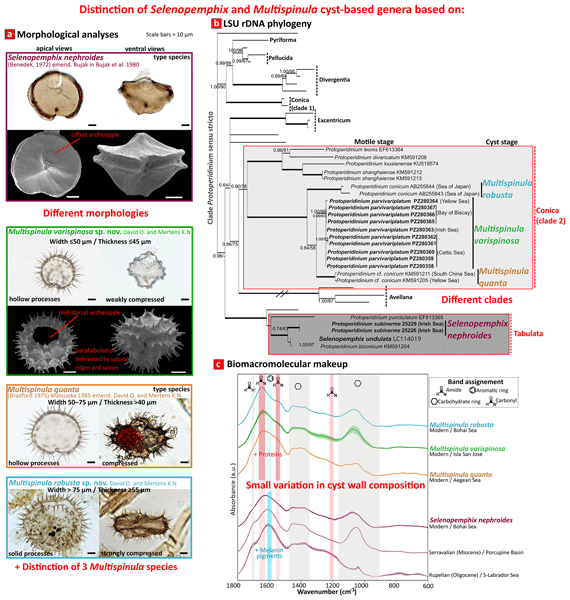
New molecular phylogenetic analyses demonstrate that the two species are polyphyletic, supporting the distinction of two cyst-based genera. Morphological features further support this distinction and justify emended diagnoses for both genera. The genus Selenopemphix is emended to include only cysts with an offset archeopyle and ornamentation restricted to the paracingular margins, while the genus Multispinula is emended to include cysts with a mid-dorsal archeopyle and paratabulation outlined by parasutural ridges and spiny processes present in the precingular and postcingular regions. Selenopemphix quanta is transferred back to its initial name Multispinula quanta. Furthermore, two new species, Multispinula robusta sp. nov. and Multispinula varispinula sp. nov., are described as cyst stages of Protoperidinium conicum and Protoperidinium parvivariplatum sp. nov. In addition, our results confirm the equivalence between S. nephroides and Protoperidinium subinerme. Cysts of modern Multispinula and Oligocene to modern Selenopemphix nephroides exhibit a proteinaceous and pigmented (melanised) wall composition, common for protoperidinioids. However, Multispinula cysts are chemically distinct from Selenopemphix cysts in being relatively less melanised and more enriched in proteins. High protein abundances in dinocyst walls are evaluated to negatively affect preservation potential.
This study stabilises the taxonomy of the cyst genera Multispinula and Selenopemphix and highlights the value of integrating morphological, molecular, and cyst wall chemical analyses to resolve issues in dinoflagellate cyst taxonomy.
- Article
(15877 KB) - Full-text XML
-
Supplement
(1335 KB) - BibTeX
- EndNote


Dinoflagellates are unicellular and largely planktonic organisms which can produce resting cysts during their life cycle. These dormant stages often result from sexual fusion, whereupon they are termed hypnozygotic cysts. The existence of two life stages has resulted in the erection of biological and palaeontological classification systems, leading to separate names for the living (motile stage) and the fossil (resting or cyst stage) counterparts, the so-called dual nomenclature sanctioned by the International Code of Nomenclature (ICN) for algae, fungi, and plants (Head et al., 2024).
The heterotrophic thecate dinoflagellate genus Protoperidinium has a complicated taxonomic history, as summarised by several authors (Taylor, 1976; Abé, 1981; Harland, 1982). Protoperidinium species can be classified using complementary approaches: morphological studies of both the thecate and resting stage and, more recently, using molecular phylogenetics (e.g. Yamaguchi et al., 2006; Liu et al., 2014; Gu et al., 2015; Mertens et al., 2017). The thecate stage of the genus Protoperidinium is characterised by three cingular plates and an additional transitional plate (Balech, 1974). The 303 currently accepted species (Guiry and Guiry, 2018) are then distinguished based on body size, shape, presence, and position of apical/antapical horns and spines, cingular displacement, and particularly plate topology and plate shapes (Hoppenrath, 2009). Different subgenera are defined based on the number of anterior intercalary (a) and precingular plates (′′): firstly Protoperidinium (3a, 7′′), Minusculum (3a, 6′′), and Archaeperidinium (2a, 7′′) (Balech, 1974), and later Testeria (1a, 7′′ and without apical pore complex; Faust, 2006) was introduced. More recent molecular analyses have questioned the validity of the subgenus Minusculum (Yamaguchi et al., 2006; Ribeiro et al., 2010), have demonstrated the polyphyly of Protoperidinium, and reinstated the subgenus Archaeperidinium (Yamaguchi et al., 2011). Based on the shape of the first apical (1′) plate and the second anterior intercalary (2a) plate, the subgenus Protoperidinium can be subdivided into different sections (e.g. Gribble and Anderson, 2006). Molecular phylogenies indicated that most of these sections are monophyletic and embedded within the Protoperidinium sensu stricto clade (Mertens et al., 2013; Gu et al., 2015). There are exceptions, as the section Conica is polyphyletic within the Protoperidinium clade (Yamaguchi et al., 2006; Gu et al., 2015). The section Conica 2 is characterised by an ortho 1′ and a hexa 2a (comprising P. conicum, P. divaricatum, P. leonis, P. lousianense, and P. shanghaiense), whereas the section Tabulata is characterised by an ortho 1′ and a penta 2a (comprising P. biconicum, P. humile, and P. punctulatum).
Resting cysts produced by Protoperidinium display diverse morphologies and are classified into several genera. One of them, the genus Selenopemphix, comprises 29 fossil and extant species (File S1 in the Supplement), with the oldest, Selenopemphix maastrichta, dating back to the Late Cretaceous (Kumar et al., 1993). The genera Multispinula and Omanodinium are considered junior synonyms (Bujak et al., 1980; Bradford and Wall, 1984; Matsuoka, 1985; Head, 1993), whereas Margosphaera is considered a senior synonym, and Selenopemphix has been conserved against it (Head, 1993; Fensome et al., 2016). The genus Selenopemphix was erected on the basis of an Oligocene specimen of Selenopemphix nephroides to designate distinctive Cenozoic dinoflagellate cysts as being characterised by thin brown walls and kidney-shaped capsules in polar view with a deeply incised parasulcus and flattened dorso-ventrally and as having two rounded antapical protrusions (Benedek, 1972; Bujak et al., 1980). The species of the genus have a wide cingulum formed by two parallel ridges, with margins or crests that can be verrucae or crenulate or that can bear long to short bifurcate or trifurcate processes or spines. However, the position of the archeopyle, symmetric or offset relative to the mid-dorsal line, has been a subject of debate (Bujak et al., 1980; Head, 1993; Lentin and Williams, 1989; Mertens et al., 2017).
The status of Multispinula is problematic. Initially described as a separate genus, based on the type species Multispinula quanta (from recent sediments of the Persian Gulf; Bradford, 1975), it comprises proximate cysts that are circular to rhomboidal in ambitus and ovoidal to reniform in polar view. They bear rows of solid spines, lack antapical horns, and exhibit well-defined cingular and sulcal zones. Later, Selenopemphix was emended to include spinate species (Bujak et al., 1980), and Matsuoka (1985) considered Multispinula a junior synonym of Selenopemphix, following Bujak et al. (1980) and Harland (1982). This transfer was rejected by Lentin and Williams (1989) on the grounds that the archeopyle position had not been demonstrated as offset, which was then a morphological criterion for defining a Selenopemphix species, although this has been questioned by some authors (Bujak et al., 1980). The genus Selenopemphix was once again emended to include cysts with both a symmetrically located and offset archeopyle (Head, 1993), thereby validating the transfer. Lentin and Williams (1989) also argued for retaining Multispinula as a distinct genus based on spine distribution, an argument dismissed by Head (1993), who observed specimens with variable process distribution and with processes always aligned along the paracingular margins, which is a characteristic of Selenopemphix.
New phylogenies of Protoperidinium corroborate morphological classifications derived from motile stages and their harmonisation with cyst-based taxonomy (Sarai et al., 2013; Mertens et al., 2017; Gurdebeke et al., 2020). In these phylogenies, the species Protoperidinium conicum, previously related to the cyst Selenopemphix/Multispinula quanta, and the cyst-based species Selenopemphix undulata are placed into different sections of the Protoperidinium sensu stricto clade – Conica and Tabulata, respectively (Mertens et al., 2017). This polyphyly coupled with morphological differences (i.e. archeopyle position) between both cysts, as suggested by some authors (Lentin and Williams, 1976; Mertens et al., 2017), has rekindled discussions about the distinction between Selenopemphix and Multispinula. However, the phylogenetic position of the type species of Selenopemphix, Selenopemphix nephroides, needed to support the separation of these genera was lacking. In addition, molecular approaches also revealed that there might be several species in the “Protoperidinium conicum complex” (Yamaguchi and Horiguchi, 2005; Yamaguchi et al., 2006; Gu et al., 2015), which could explain the broad morphological variability (wide range in body size) observed among the cyst-species equivalent, currently grouped under Selenopemphix/Multispinula quanta (Head, 1996; Rochon et al., 1999).
The main issues addressed in this study are (i) whether Selenopemphix and Multispinula represent distinct genera and (ii) if the morphological variability observed in cysts assigned to Multispinula/Selenopemphix quanta reflects hidden diversity within the “Protoperidinium conicum complex”. To address these issues, we investigate the morpho-molecular relationships between the respective type species – Selenopemphix nephroides and Multispinula quanta – through a combination of incubation experiments on cysts extracted from surface sediments (to establish cyst–theca relationships), detailed morphological observations using light microscopy (LM) and scanning electron microscopy (SEM), and molecular analyses based on large-subunit (LSU) ribosomal DNA (rDNA) sequences. Molecular markers are used to infer evolutionary relationships, detect cryptic diversity, and better assess species boundaries in morphologically similar dinoflagellates. In addition, we apply single-specimen attenuated total reflection Fourier transform infrared microspectroscopy (ATR µ-FTIR) to characterise the biomacromolecules comprising cyst walls, allowing assessment of inter-taxon compositional variability and preservation potential in the fossil record. The integration of morpho-molecular and FTIR-based cyst wall chemical approaches has been used for roughly a decade and provides a more holistic perspective to resolve taxonomic issues in modern and fossil dinocysts (e.g. Mertens et al., 2017, 2023, 2024; Gurdebeke et al., 2020).
2.1 Sediment samples and germination experiments
Surface sediments containing living dinoflagellate cysts were collected from several locations in the Atlantic Ocean (Celtic Sea, Irish Sea, Bay of Biscay) and in the Pacific Ocean (Yellow Sea, Lake Saroma) (Table 1). After sampling, materials were stored in plastic containers in the dark at 4 °C until further treatment.
For the sample from Lake Saroma, the >150 µm sediment fraction was processed using hydrochloric acid (HCl) at room temperature and warm hydrofluoric acid (HF) to remove the mineral fraction and concentrate cysts. For the other samples studied, approximately 2–5 g of wet sediment mixed with filtered seawater was ultrasonicated for 5 min and rinsed through a 125 and 20 µm metallic-meshed sieve. From this residue, the cyst fraction was separated using the heavy liquid sodium polytungstate (SPT; density = 1.4 g cm−3) method (Bolch, 1997). For cyst incubation experiments, single cysts were isolated with a micropipette using an inverted light microscope (Olympus IX70). Then, they were individually transferred into 0.5 mL microwells filled with L1 medium and incubated under the following conditions: 16 °C, 12 h light : 12 h dark photocycle. The incubated cysts were regularly checked for germination.
Table 1Locations of investigated surface sediment samples, along with identified species (dinocysts and their corresponding germinated cells observed in this study) and associated LSU rDNA sequence codes.

2.2 Microscopy observations
Light microscopy (LM) observations were conducted on (empty and living) cysts and germinated cells, using an Olympus IX-TVAD (Japan) camera mounted on an Olympus IX70 microscope or on an Olympus BX41 microscope equipped with 100× oil immersion objective. To study the shapes of thecal plates, vegetative cells that hatched from cysts were transferred onto glass slides, stained using solophenyl flavine 7GFE 500 (Ciba Specialty Chemicals, High Point, North Carolina USA; Chomérat et al., 2017), and observed under the Olympus BX41 equipped with a U-MWU2 Olympus fluorescent filter cube.
For scanning electron microscopy (SEM), isolated cysts and cells were transferred onto polycarbonate membrane filters (Isopore membrane filter, 0.5 µm pore size) using a micropipette and processed according to Chomérat and Couté (2008). Dried membranes were affixed to aluminium stubs with adhesive tabs (Electron Microscopy Sciences, Hatfield, Pennsylvania, USA) and coated for 60 s with gold using a Cressington 108Auto sputter coater. Observations were carried out at the Station of Marine Biology in Concarneau with a Zeiss Sigma 300 field-emission SEM equipped with a conventional Everhart–Thornley detector and in-lens detectors of secondary electrons at 1.5 and 5 kV.
Cyst and cell dimensions were measured from SEM or LM images using ImageJ software (Rasband, 1997). For each motile cell, length and width were measured. For each cyst, the longest and shortest body diameters and five processes were measured. All motile cell measurements in the species descriptions cite the minimum, average (in parentheses), and maximum values (in µm), in that order. The Kofoidian system was used for labelling tabulations; sulcal plate labels are in accordance with Balech (1980).
2.3 Molecular and phylogenetic analyses
Single-cell PCR amplification and sequencing. For North Atlantic samples, germinated single cells or living cysts were isolated; cysts were mechanically disrupted by gentle squeezing. Cells were rinsed several times in sterile distilled water, transferred into 0.2 mL polymerase chain reaction (PCR) tubes, and preserved at −20 °C until analysis. LSU rDNA fragments were amplified using a nested PCR approach. PCR reactions were performed in a final volume of 25 µL using Promega Master Mix (Promega, Madison, USA), following the manufacturer's instructions. In the first PCR round, the primers 515F (Turner et al., 1999) and D3B (Nunn et al., 1996) were used to amplify the gene coding for the LSU rRNA. A second round of PCR was conducted using 1 µL of the first-round amplicon as a template and the primer pairs D1R (Scholin, 1994) and D3B. Amplifications were conducted in a thermal cycler (Biometra Trio, Analytik Jena) under the following cycling conditions: an initial denaturation step of 2 min at 94 °C, 35 cycles at 94 °C for 30 s, annealing at 60 °C for PCR1 or 56 °C for PCR2 for 1 min, and an extension at 72 °C for 3 min 30 s. This was followed by a final extension at 72 °C for 5 min. The amplified products were visualised on a 1 % agarose gel after electrophoresis, and positive amplicons were purified using the ExoProStar DNA purification kit (Cytiva, USA). Purified products were then sent to Microsynth (France) for Sanger sequencing using the primers from the second PCR round. Both forward and reverse reads were obtained.
For the Yellow Sea sample, the germinated single cell was used as the template to amplify about 1430 bp of the nuclear-encoded LSU rDNA (D1–D6 domains) using the primers D1R and 28-1483R (Daugbjerg et al., 2000). A 50 µL PCR cocktail was prepared containing 0.2 µM of both forward and reverse primers, 5 µL of 20 mM Ex Taq buffer, 4 µL of a 2.5 mM dNTP mixture, and 0.25 µL of Ex Taq DNA polymerase (5 U µL−1, Takara, Dalian, China). The final volume was adjusted to 50 µL using sterilised distilled water. Thermal cycling was conducted using a Mastercycler PCR system (Eppendorf, Hamburg, Germany) under the following cycling steps: an initial denaturation step of 3.5 min at 94 °C, 35 cycles at 94 °C for 50 s, annealing at 45 °C for 50 s, an extension at 72 °C for 80 s, and a final extension of 10 min at 72 °C. The PCR products were sequenced in both directions using the ABI BigDye dye-terminator technique (Applied Biosystems, Foster City, CA, USA) according to the manufacturer's instructions.
Sequence alignments and phylogenetic analyses. Newly obtained sequences were first aligned with those of related species available in GenBank using the Mafft algorithm with the selection of the q-ins-i strategy (Katoh et al., 2019) and were subsequently cut. Akashiwo sanguinea (Hirasaka) G. Hansen & Moestrup, in Daugbjerg et al. (2000), was selected as the outgroup. Two methods of phylogenetic reconstruction were used in which a general time-reversible model (GTR + I + G) was selected. A Bayesian reconstruction of the data matrix was performed with MrBayes 3.2.7 (Ronquist et al., 2012). Four Markov chain Monte Carlo (MCMC) chains ran for 1 million generations, sampling every 100 generations with a burn-in of 10 %. Maximum likelihood (ML)-based analyses were conducted using Mega12 (Kumar et al., 2024). Around 10 000 bootstrap replicates were performed to assess the relative robustness of branches of the ML tree. Statistical support values (ML bootstrap support, Bayesian posterior probabilities) were drawn on the resulting best-scoring tree.
2.4 Macromolecular characterisation of the cyst wall
Cyst wall biomacromolecules were analysed via attenuated total reflection Fourier transform infrared microspectroscopy (ATR µ-FTIR) using the protocol of Meyvisch et al. (2022). Prior to analysis, individual dinocysts were isolated from a variety of modern and fossil sediment samples processed with different extraction methods to yield concentrated organic residues in distilled water (Table 2). Isolation from residue droplets was performed under a Zeiss Primovert inverted microscope (Carl Zeiss AG) at 100× or 200× magnification using a narrowed glass Pasteur pipette attached to a rubber suction tube. Each chosen specimen was photographed at 400× magnification with an AxioCam MRc5 (Carl Zeiss AG) and subsequently deposited onto a pre-labelled gold-coated mirror (Thorlabs; model: PFSQ20-03-M03). This mirror was transferred to the stage of a Hyperion 2000 microscope coupled to a Bruker Vertex 80v FTIR spectrometer (Bruker Corporation), and individual specimens were analysed after being brought in contact with a Ge micro-ATR objective. Spectra were collected using a liquid-nitrogen-cooled mercury cadmium telluride detector over the range of 4500–600 cm−1 at a spectral resolution of 4 cm−1, with 256 scans averaged per spectrum. Atmospheric CO2 and H2O contributions were removed from the raw data using OPUS 8.2.21 software (Bruker Corporation). The processed spectra were individually exported as *.txt files and combined with associated metadata into a single dataset (File S2). This dataset was further processed using the “Preprocess Spectra” widget in Quasar 1.7.0 open-source software Toplak et al., 2021). Processing included Savitzky–Golay smoothing (window size = 9, polynomial order = 2, derivative order = 0), truncation to 3800–600 cm−1, rubber band baseline correction, and vector normalisation, in that order. Spectral plots were exported from Quasar as *.svg files and further edited in Inkscape v.1.3.2 (https://inkscape.org). The identification of spectral absorption bands was based on Coates (2000) and Meyvisch et al. (2023).
Table 2Information on samples and extracted dinocysts used for biomacromolecular analyses (ATR µ-FTIR). SS: short spines. SL: short and long spines. LS: long spines. SPT: sodium polytungstate. HCl: hydrochloric acid. HF: hydrofluoric acid.

3.1 Results of germination experiments and microscopy observations
Several cysts of Protoperidinium were isolated from surface sediments of the Atlantic Ocean (Celtic Sea, Irish Sea, Bay of Biscay) and the Pacific Ocean (Yellow Sea, Lake Saroma; Table 1). Thirty-nine cysts germinated into motile cells. Five cells of Protoperidinium subinerme emerged from the cyst species Selenopemphix nephroides collected from the Irish Sea. Twenty-nine cells were identified here as Protoperidinium parvivariplatum sp. nov., equivalent to the new cyst-defined species Multispinula varispinosa sp. nov. Five cells germinated from the cyst Multispinula quanta and were identified as Protoperidinium cf. conicum. Finally, cysts of Multispinula robusta sp. nov. were identified from the palynologically treated Lake Saroma sample.
3.2 Systematic part
-
Division DINOFLAGELLATA (Bütschli) Fensome et al., emend. Adl et al.
-
Class DINOPHYCEAE Pascher
-
Subclass PERIDINIPHYCIDAE Fensome et al.
-
Order PERIDINIALES Haeckel
-
Family PROTOPERIDINIACEAE Balech nom. cons.
-
Subfamily PROTOPERIDINIOIDEAE (autonym)
-
Genus Protoperidinium Bergh
-
Protoperidinium subinerme (Paulsen) Loeblich III Plate 1, figs. 1–14

Plate 1LM and SEM micrographs of Protoperidinium subinerme (1–14) hatched from Selenopemphix nephroides (15–21). Specimens are from the Irish Sea (United Kingdom, station ST5: 53°19′6.4′′ N, 3°06′42.3′′ W). Scale bars = 10 µm. (1) Mid-focus on a living cell in ventral view showing the body shape and cell contents. (2–5) Fluorescence micrographs of a stained cell, in high focus: ventral-apical view showing an ortho first apical plate (1′) (2), dorso-apical views showing a hexa second anterior intercalary plate (2a) (3–4), focus on two short antapical protrusions highlighted by two white arrows (5). (6–9) SEM micrographs of a thecate: ventral view (6), focus on the apical pore complex (7), apical view (8), and focus on two short antapical protrusions highlighted by two white arrows (9). (10–14) SEM micrographs of a thecate: antapical (10), ventral (11), left-antapical (12), right-antapical (13) views and focus on the sulcal area (14). (15) Living cyst with cell contents: mid-focus in ventral view. Grey arrows indicate the two weakly developed horns. (16) Living cyst with cell contents: mid-focus in polar view. (17–18) Empty cyst: high-focus in ventral view (17) and mid-focus in polar view (18). Black arrows indicate one of the flagellar scars. (19–21) SEM micrographs of a living cyst with a preformed archeopyle: polar (19) and ventral (20) views and focus on the archeopyle (21). Grey arrows indicate the two weakly developed horns.
Description. The motile cells hatched from cysts of the Irish Sea and are rhombic in ventral view (Plate 1, figs. 1, 6). Both epi- and hypo-theca have convex sides. The plate tabulation is Po, X, 4′, 3a, 7′′, 3C+t, 4S, 5′′′, 2′′′′ (Plate 1, figs. 6–14). The apical pore complex is composed of an oval apical pore plate (Po), surrounded by raised ridges of the surrounding apical plates, and by a short and trapezoidal canal plate (X) (Plate 1, fig. 7). The first apical plate (1′) is quadrangular (ortho-type) and symmetrical (Plate 1, figs. 2, 6, 8). Apical plates 4′ and 2′ are hexagonal, whereas plate 3′ is pentagonal (Plate 1, fig. 8). There are three anterior intercalary plates; plates 1a and 3a are pentagonal, whereas 2a is hexagonal and deltaform-linteloid (Plate 1, figs. 3–4, 8). The epitheca is asymmetrical, the 2a plate appears offset left of the dorsal midline (Plate 1, fig. 8). The cingulum located in the equatorial part of the cell is slightly descending (laevorotatory), from one cingulum width. There are at least four sulcal plates (Plate 1, figs. 6, 11, 13–14). The anterior sulcal plate (Sa) is short and contacts 1′, 1′′, and 7′′ anteriorly. The right sulcal plate (Sd) is subrectangular and anteriorly connected to plate 7′′ through a long and narrow protrusion in its anterior part (Plate 1, fig. 11). The left sulcal plate (Ss) is narrow and subrectangular, and the posterior sulcal plate (Sp) is large-sized and U-shaped, connected to plates 5′′′, 2′′′′, 1′′′, and 1′′′′ (Plate 1, fig. 6, 13–14). All postcingular plates are quadrangular, except for 3′′′, which is pentagonal. The two antapical plates (1′′′′ and 2′′′′) are pentagonal and similar in size (Plate 1, fig. 10). Two or more protrusions are present on the antapical plates (Plate 1, figs. 5, 9). Peripheral cell contents are pinkish and yellowish.
Dimensions. Cells germinated from incubated cysts from the Irish Sea (western England coast) measure 47.1(50.5)52.4 µm in length and 46.8(51.1)57.9 µm in width (N=5). Detailed measurements of the observed cells are provided in File S3.
Gene sequence. The LSU rDNA gene sequence of the cell germinated from a cyst collected in sediments of the Irish Sea with GenBank accession no. PZ280369. The LSU rDNA gene sequence of the cyst collected in the surface sediment of the Irish Sea with GenBank accession no. PZ280368.
Equivalent. Protoperidinium subinerme is related to the cyst species Selenopemphix nephroides, as confirmed by incubation experiments performed in the present study.
-
Protoperidinium parvivariplatum sp. nov. David O. et Mertens K.N.
Plate 2, figs. 1–17

Plate 2LM and SEM micrographs of Protoperidinium parvivariplatum sp. nov. hatched from Multispinula varispinosa sp. nov. illustrated in Plates 4, 5, and 6. Specimens are from the Celtic Sea (Ireland, station CP4: 51°44′13.2′′ N, 6°22′19.2′′ W) and the Bay of Biscay (France, station Scoré: 47°52′31.7′′ N, 3°57′15.1′′ W). Scale bars = 10 µm. (1) Mid-focus on a living cell (station CP4) showing the body shape and cell contents. The cell germinated from a long-spined cyst as illustrated in Plate 5. (2–4) Fluorescence micrographs of a stained cell (station CP4), in high focus: ventral view showing an ortho first apical plate (1′) (2), dorsal views showing a hexa second anterior intercalary plate (2a) (3–4). (5) Mid-focus on a living cell (station CP4) showing the body shape and cell contents. The cell germinated from a short-spined cyst as illustrated in Plate 6. (6–12) Fluorescence micrographs of a stained cell (station CP4), in high focus: ventral-apical view showing an ortho first apical plate (1′) (6); dorso-apical views showing a dextro-penta second anterior intercalary plate (2a) (7–8); right-antapical (9), left-antapical (10), ventral-antapical (11), and dorso-antapical (12) views. (13–17) Holotype – SEM micrographs of a thecate (station Scoré): ventral (13–14) and antapical (15) views, focus on the apical pore complex (16) and the sulcus (17).
Derivation of name. The epithet reflects the small size of the cell (from Latin: parvus, meaning small) and the variability in the anterior intercalary plate 2a (from Latin: variplatum, meaning variable plate).
Diagnosis. The motile cell is small and pentagonal, with plate formula Po, X, 4′, 3a, 7′′, 3C+t, 4S, 5′′′, 2′′′′. The epitheca have straight to slightly convex sides. The first apical plate (1′) is large, quadrangular (ortho-type), and symmetrical. The second anterior intercalary plate (2a) is quadra, sinistro-/dextro-penta or hexa and deltaform-linteloid. The cingulum shows no displacement. The motile cells show a short apical horn and two acuminate antapical horns of equal length or with one slightly shorter, each bearing a short spine. All thecal plates are reticulated with trichocyst pores positioned on the sutures of the reticulations, except for APC + X.
Holotype. Specimen shown in Plate 2, figs. 9–13. The specimen illustrated is on a SEM stub (designated CEDiT2026H208) curated at the Senckenberg Research Institute and Natural History Museum, Centre of Excellence for Dinophyte Taxonomy, Germany.
Type locality. Concarneau Bay (southern Brittany coast, north-western France), northern part of the Bay of Biscay (station Scoré at 47°52′31.7′′ N, 3°57′15.1′′ W).
Description. The motile cells hatched from cysts from different localities (Irish Sea, Celtic Sea, Bay of Biscay, Yellow Sea) are small and pentagonal in outline and carry an apical horn and two antapical horns of equal length or with one appearing shorter, each bearing a short spine. The epitheca is conical in ventral view with straight or very slightly convex lateral margins (Plate 2, figs. 1, 5). The cingulum is located in the equatorial part of the cell and does not show displacement. The sutures that run from the apex to the cingulum are not completely straight (Plate 2, figs. 6, 14). The thin thecal plates carry polygonal reticulations with trichocyst pores positioned on the sutures of the reticulations, except for APC + X (Plate 2, figs. 7–17). The plate tabulation is Po, X, 4′, 3a, 7′′, 3C+t, 4S, 5′′′, 2′′′′. The apical pore complex is surrounded by a very low apical collar formed by the raised edges of apical plates 2′ and 4′; it is composed of an oval apical pore plate (Po) and an elongate and trapezoidal canal plate (X) (Plate 2, fig. 16). The first apical plate (1′) is wide, symmetrical, and rhombic (ortho-type) (Plate 2, figs. 2, 6, 13). Plates 2′ and 4′ are elongated and quadra/pentagonal and pentagonal, respectively (Plate 2, figs. 6, 9–10, 14). Plate 3′ is compact and pentagonal (Plate 2, fig. 7). There are three anterior intercalary plates (1a, 2a, and 3a), and plates 1a and 3a are penta/hexa (Plate 2, figs. 9–10). The second intercalary plate (2a) displays various morphologies: quadra, sinistro-/dextro-penta, or hexa and deltaform-linteloid (Plate 2, figs. 3–4, 7–8). The precingular series consist of seven plates. Plates 1′′ and 7′′ are triangular or quadrangular, whereas 2′′, 3′′, 5′′, and 6′′ are quadrangular (Plate 2, figs. 9–10, 14). Plate 4′′ is quadrangular when 2a is quadra or hexa and penta when 2a is penta (Plate 2, figs. 3–4, 7–8). There are three cingular plates plus a transitional plate (t). There are four sulcal plates (Plate 2, figs. 13, 15, 17). The anterior sulcal plate (Sa) is short and subrectangular, with its anterior part contacting 1′ and slightly 1′′ but not 7′′. The right sulcal plate (Sd) is long and narrow, contacting the 7′′ plate through a long protrusion in its anterior part. The left sulcal plate (Ss) is long and formed a J-shaped curve (Plate 2, fig. 15). The posterior sulcal plate (Sp) is long and asymmetrically U-shaped, contacting plates 5′′′ and 2′′′′ and plates 1′′′ and 1′′′′ through a long and narrow extension. Postcingular plates 1′′′, 2′′′, 3 ′′′, and 5′′′ are pentagonal, whereas 2′′′ and 4′′′ are quadrangular. The two antapical plates (1′′′′, 2′′′′) of similar size are pentagonal and formed the antapical horns. Peripheral cell contents are pinkish.
Dimensions. The holotype measures 34.7 µm in length and 41.6 µm in width. The cingular width measures 3.9 µm, and the distance between the two antapical horns is 16 µm. The other cells observed in this study measure 38.6(49.6)59.0 µm in length, 38.0(46.2)55.0 µm in width (N=25). Detailed measurements of the observed cells are provided in File S3.
Gene sequence. The LSU rDNA gene sequence of the cells germinated from cysts collected in sediments of the Celtic Sea with GenBank accession nos. PZ280358, PZ280359, PZ280360, PZ280361, and PZ280362. The LSU rDNA gene sequence of the cell germinated from a cyst collected in sediments of the Irish Sea with GenBank accession no. PZ280363. The LSU rDNA gene sequence of the cyst collected in sediments of the Yellow Sea with GenBank accession no. PZ280364. The LSU rDNA gene sequence of the cysts collected in sediments of the Bay of Biscay with GenBank accession nos. PZ280365, PZ280366, and PZ280367.
Equivalent. Protoperidinium parvivariplatum is related to the cyst species Multispinula varispinosa, as shown by incubation experiments performed in the present study.
Remarks. Protoperidinium conicum described from the Norwegian coast differs by its larger size (70 µm in length, 75 µm in width), its epitheca with straight to concave sides, and its hexa 2a plate. Protoperidinium cf. conicum appears quite similar but differs by its larger size (60–66 µm in length, 50–56.8 µm in width), its epitheca with concave sides, and its hexa to quadra 2a plate.
-
Protoperidinium cf. conicum
Plate 3, figs. 1–7

Plate 3LM micrographs of Protoperidinium cf. conicum (1–7) hatched from Multispinula quanta (8–15). Specimens are from the Celtic Sea (Ireland, station CP4: 51°44′13.2′′ N, 6°22′19.2′′ W; Ireland, station DN1: 52°3′18.0′′ N, 7°29′49.2′′ W; Ireland, station DN3: 51°41′49.2′′ N, 7°31′19.2′′ W) and the Bay of Biscay (France, station Scoré: 47°52′31.7′′ N, 3°57′15.1′′ W). Scale bars = 10 µm. (1) Mid-focus on a living cell (station DN1) showing the body shape and cell contents. (2–7) Fluorescence micrographs of a stained cell (station CP4), in high focus: ventral-apical view showing an ortho first apical plate (1′) (2); dorso-apical views showing a hexa second anterior intercalary plate (2a) (3); right-antapical (4), left-antapical (5), ventral-antapical (6), and dorso-antapical (7) views. (8–9) Living cyst with cell contents (station CP4): high-focus in dorsal views (8) and mid-focus in polar view (9). (10–12) Living cyst with cell contents (station Scoré): mid-focus (10) to high-focus (11) in dorsal view and high-focus in polar view (12). (13–15) Empty cyst (station DN3): mid-focus (13) to high-focus (14) in dorsal view and mid-focus in polar view (15). Black arrow indicates one of the flagellar scars.
Description. The motile cells are large and pentagonal, with plate formula Po, X, 4′, 3a, 7′′, 3C+t, ?S, 5′′′, 2′′′′ (Plate 3, figs. 1–7). These cells carry an apical horn and two long antapical horns of equal length, each bearing a short spine. The epitheca has concave sides. The sutures that run from the apex to the cingulum are almost straight (Plate 3, fig. 2). The cingulum is located in the equatorial part of the cell and does not show displacement. The first apical plate (1′) is symmetrical, elongated, and rhombic (ortho-type) (Plate 3, fig. 2). Plate 2′, 3′, and 4′ are pentagonal. There are three anterior intercalary plates, with the second intercalary plate (2a) being quadrangular (Plate 3, fig. 3). The thin thecal plates carry spiny reticulations (Dodge, 1983; Plate 3, fig. 4).
Dimensions. The cells measure 63.0(73.5)84.0 µm in length and 57.0(65.5)76.0 µm in width (N=5). Detailed measurements of the observed cells are provided in File S3.
Equivalent. Protoperidinium cf. conicum is related to the cyst species Multispinula quanta, as shown by incubation experiments performed by Gu et al. (2015) and in the present study.
Remarks. Protoperidinium conicum described from the Norwegian coast differs by its larger size (70 µm in length, 75 µm in width), its epitheca with straight to concave sides, and its hexa 2a plate. Protoperidinium parvivariplatum appears quite similar but differs by its smaller size (38.8–59 µm in length, 36.6–55 µm in width), its epitheca with convex sides, and its variable 2a plate (quadra, penta, hexa).
-
Genus Selenopemphix Benedek 1972 emend. nov. David O. et Mertens K.N.
Synonym.
-
1968 ?Margosphaera Nagy, p. 208, table V, figs. 23–25.
-
1972 Selenopemphix Benedek, pp. 47–48, pl. 11, fig. 13, and pl. 16, figs. 1–4.
-
1975 Omanodinium Bradford, pp. 3070–3074, figs. 23–28.
-
1980 Selenopemphix (Benedek, 1972) emend. Bujak in Bujak et al., 1980, pp. 82–83.
-
1993 Selenopemphix (Benedek, 1972) emend. Bujak in Bujak et al., 1980, emend. Head, pp. 32–34, fig. 20.
Type species. Selenopemphix nephroides Benedek, 1972, pp. 47–48, pl. 11, fig. 13.
Original diagnosis. (Benedek, 1972, p. 47) Thin-walled dorso-ventrally flattened shell without apical process but with two antapical prominences. The outer wall of the shell is weakly granular and generally somewhat folded. A trapezoidal opening pierces the apical area laterally.
Emended diagnosis. Polar compressed peridinioid cyst with an ovoidal to reniform outline in polar view. In dorsal-ventral view, outline is broadly peridinioid, pentagonal, or rhomboidal. The wall is single-layered and brown to light brown in colour. The wall surface is smooth, shagreenate, granulate, or perforate. The paracingulum is excavated and planar or weakly descending. The paracingular margins are more or less raised; can bear crests, grana, verrucae, processes, or spines; and are interrupted by the parasulcus. Ornamentations, when present, are restricted to the paracingular margins, only rarely occur on the apical and antapical horns, and are consistently absent from the precingular and postcingular zones. No paratabulation is visible, with the exception of the paracingulum, the parasulcus, and the archeopyle. The archeopyle is formed by loss of the 2a anterior intercalary paraplate, is offset relative to the mid-dorsal line, and has rounded angles. The operculum is free or remains adherent.
Remarks. The genus Omanodinium Bradford, which was described with the type species Omanodinium alticinctum (originating from recent sediment of the Oman Gulf), is considered to be a taxonomic junior synonym of Selenopemphix (Bradford and Wall, 1984; Head, 1993). In addition, the monospecific acritarch genus Margosphaera Nagy, with its type Margosphaera velata described from the Miocene of Hungary, is considered to be a taxonomic senior synonym of the genus Selenopemphix, considering the similar morphology between S. nephroides and M. velata, based on the re-examination of published photomicrographs (Fensome et al., 2016; Head, 1993). The genus Multispinula Bradford, which was erected with the species Multispinula quanta, was later considered to be a junior synonym of the genus Selenopemphix by Head (1993), who emended Selenopemphix to accommodate cysts with both symmetrically located and offset archeopyles. The genus Selenopemphix is emended here to exclude species with symmetrically located archeopyles and cysts presenting an ornamentation (spines) on the precingular and postcingular zones, which are considered one of the main characteristic features of the genus Multispinula.
-
Selenopemphix nephroides (Benedek 1972) emend. Bujak in Bujak et al. 1980
Plate 1, figs. 15–21
Description. Cysts extracted from surface sediment of the Irish Sea are reniform in polar view, showing a polar compression (Plate 1, figs. 16, 18–19). The wall is light to medium brown and fairly smooth. The epicyst is ornamented with faint striations that run from the apex to the paracingulum (Plate 1, figs. 17, 20). The deeply excavated, descending (approximately of cingular width) paracingulum divides the epicyst and hypocyst, with the hypocyst being slightly longer (Plate 1, figs. 15, 17, 20). The paracingular margins are denoted by entirely distal, raised ridges with occasional notches and are absent above the shallow parasulcus (Plate 1, figs. 16, 18, 19). The epicyst and hypocyst have concave and convex sides, respectively (Plate 1, fig. 20). The epicyst displays a small, rounded horn. Distally, the hypocyst expresses two weakly developed rounded horns, positioned close to each other (Plate 1, figs. 15, 20). Two flagellar scars are present just below the paracingulum. No paratabulation pattern is observed, with the exception of the archeopyle, the paracingulum, and the parasulcus. The archeopyle is formed by the loss of the 2a anterior intercalary paraplate and is deltaform-linteloid with rounded angles and offset to the left of the dorsal midline (Plate 1, figs. 19–20). It results from the partial or complete detachment of the second anterior intercalary paraplate 2a. The operculum is free.
Dimensions. Cysts collected from surface sediments of the Irish Sea used for hatching experiments measure 37.2(44.9)48.3 µm in length, 51.7(63.2)71.0 µm in width, and 50.0(57.9)63.8 µm in thickness (N=14). Detailed measurements of the observed cysts are provided in File S3.
Stratigraphic range. Lower Eocene (De Coninck, 1977; King et al., 2018) to recent (Thöle et al., 2023; this study).
Gene sequence. The LSU rDNA gene sequence of the cyst collected in the surface sediment of the Irish Sea with GenBank accession no. PZ280368. LSU rDNA gene sequence of the cell germinated from a cyst collected in sediments of the Irish Sea with GenBank accession no. PZ280369.
Equivalent. Selenopemphix nephroides is related to Protoperidinium subinerme, according to Rochon et al. (1999) and as confirmed by incubation experiments performed in the present study.
Remarks. This species is distinguished from Selenopemphix undulata by its smooth cyst wall and the absence of undulate cingular margins (Verleye et al., 2011). The cyst of Protoperidinium biconicum is distinguished by its single blunt antapical horn (Gu et al., 2015). Selenopemphix alticincta is smaller (36 µm length, 39 µm breadth; Bradford, 1975) than S. nephroides.
-
?Selenopemphix tholus (Bradford, 1975) Head, 1996
Synonym.
-
1975 Omanodinium tholus sp. nov. Bradford, pp. 3072–3074, figs. 17–22.
-
1996 Selenopemphix tholus (Bradford, 1975) Head, p. 1231.
-
2020 Selenopemphix tholus (Bradford, 1975) Head, 1996; Mertens et al., p. 28, pl. 15, figs. 10–12.
Holotype. Omanodinium tholus Bradford 1975, p. 3073, figs. 17–18.
Stratigraphic range. Upper Oligocene (e.g. Palamarczuk and Barreda, 2000) to recent (e.g. Bradford, 1975; Mertens et al., 2020).
Remarks. This cyst exhibits a pentagonal ambitus and is circular to reniform in polar view but is not compressed apically–antapically. It also bears one apical horn and two antapical horns. These morphological features distinguish ?Selenopemphix tholus from the genus Selenopemphix. Its bilateral symmetry and pentagonal ambitus suggest a possible affinity with the genus Lejeunecysta, but a more detailed morphological analysis is necessary to confirm this hypothesis.
-
?Selenopemphix hamanaensis Kojima, 1989
Holotype. Selenopemphix hamanaensis Kojima, 1989, pp. 208–209, fig.5.
Stratigraphic range. Holocene (Kojima, 1989).
Remarks. This cyst does not exhibit the polar compression or the peridinioid shape characteristic of the genus Selenopemphix, and the archeopyle has not been observed. We therefore suggest that this specimen may represent a copepod egg, but further analyses are necessary to confirm this hypothesis.
-
Genus Multispinula Bradford 1975 emend. nov. David O. et Mertens K.N.
-
1975 Multispinula Bradford, p. 3067, figs. 5–7.
-
1993 Selenopemphix Head, pp. 31–32, figs. 20.16–22.
Type species. Multispinula quanta Bradford, 1975, p. 3068, fig. 5.
Original diagnosis. (Bradford, 1975, p. 3067) Proximate cysts: ovoidal, circular, or rhomboidal in ambitus; ovoidal to reniform in polar view. Characteristic rows of apparently solid spines ornament the phragma. No antapical horns, but with or without an apical projection. Intercalary archeopyle. No tabulation but can have a cingular and sulcal zones.
Emended diagnosis. Weakly to strongly polar compressed peridinioid cyst with an ovoidal to reniform outline in polar view. In dorsal view, the outline is broadly peridinioid, pentagonal, or rhomboidal. The wall is single-layered. The wall surface is smooth. The paracingulum is excavated and planar or weakly ascending. The paracingular margins are slightly raised and bear two parallel rows of solid to hollow spines. On the precingular and postcingular zones, other rows of spines are observed. A paratabulation is visible on the cyst, demarcated by process distribution and parasutural ridges. The archeopyle is formed by loss of the 2a anterior intercalary paraplate; has rounded angles; and is positioned on the mid-dorsal line, showing no offset. The operculum is free.
Remarks. The genus Multispinula Bradford was considered to be a junior synonym of the genus Selenopemphix by Head (1993), who emended the Selenopemphix to accommodate cysts with both symmetrically located and offset archeopyles. The genus Multispinula is emended here to encompass species bearing symmetrically positioned archeopyles and presenting a paratabulation on precingular and postcingular zones, demarcated by processes and parasutural ridges, diagnostic morphological characters that are absent from cyst species belonging to the genus Selenopemphix.
-
Multispinula varispinosa sp. nov. David O. et Mertens K.N.
Plate 4, figs. 1–11/Plate 5, figs. 1–10/Plate 6, figs. 1–10
-
1968 cyst of Protoperidinium? nudum (Meunier, 1919) sensu Wall & Dale, plate 4, figs. 1–5, pp. 277–278.
-
1997 Protoperidinium sp. 2 Sonneman & Hill, figs. 32a–d, p. 168.
-
1999 cyst of Protoperidinium nudum (Meunier, 1919) Balech 1974; Rochon et al., plate 11, figs. 5–10, p. 48.

Plate 4LM and SEM micrographs of Multispinula varispinosa sp. nov. showing long-sized spines from which Protoperidinium parvivariplatum hatched, as illustrated in Plate 2. Specimens are from the Celtic Sea (Ireland, station CP4: 51°44′13.2′′ N, 6°22′19.2′′ W). Scale bars = 10 µm. (1–2) Living cyst with cell contents: mid-focus in ventral (1) and polar (2) views. (3–4) Empty cyst: mid-focus in equatorial (3) and polar (4) views. Black arrow indicates one of the flagellar scars. (5–8) Empty cyst: mid-focus (5) to low-focus (6) in polar view and mid-focus (7) to high-focus (8) in dorso-apical view. Black arrow indicates one of the flagellar scars. (9–11) SEM micrographs of an empty cyst: antapical (9) and dorsal (10) views and focus on the long processes (11).

Plate 5LM and SEM micrographs of Multispinula varispinosa sp. nov. showing short spines from which Protoperidinium parvivariplatum hatched, as illustrated in Plate 2. Specimens are from the Celtic Sea (Ireland, station CP4: 51°44′13.2′′ N, 6°22′19.2′′ W). Scale bars = 10 µm. (1–2) Empty cyst: mid-focus (1) to high-focus (2) in dorsal view. (3–4) Mid-focus of an empty cyst: polar (3) and dorsal (4) views. Black arrow indicates one of the flagellar scars. (5) Living cyst with cell contents: mid-focus in dorsal view. (6–10) Holotype – SEM micrographs of an empty cyst: ventral (6), apical (7), left-apical (8), and right-equatorial (9) views and focus on the short processes (10).

Plate 6LM and SEM micrographs of Multispinula varispinosa sp. nov. showing long and short spines from which Protoperidinium parvivariplatum hatched, as illustrated in Plate 2. Specimens are from the Celtic Sea (Ireland, station CP4: 51°44′13.2′′ N, 6°22′19.2′′ W) and the Bay of Biscay (France, station Scoré: 47°52′31.7′′ N, 3°57′15.1′′ W). Scale bars = 10 µm. (1–2) Living cyst with cell contents (station CP4): mid-focus (1) to high-focus (2) in apical view. (3) Living cyst with cell contents (station CP4): mid-focus in polar view. (4) Living cyst with cell contents (station CP4): mid-focus in equatorial view. (5–8) SEM micrographs of an empty cyst (station CP4): antapical ventral (5), antapical dorsal (6), left-antapical (7), and right-antapical (8) views. (9–10) SEM micrographs of a living cyst with preformed archeopyle (station Scoré) ventral (9) and apical (10) views.
Derivation of name. The epithet reflects the variability in the size of the processes (from Latin vari, meaning variable, and spinosa, meaning spiny).
Diagnosis. The small-sized (width: ≤ 50 µm; thickness: ≤ 45 µm) brown cyst is reniform in polar view with a shallow parasulcus and appears weakly compressed anteroposteriorly in equatorial view. The cyst surface is smooth, ornamented by several rows of hollow spines expanded at their base. This species exhibits a wide variation in spine length; specimens could bear long spines (5–14 µm), short spines (2–4 µm), or both (3–10 µm). A paratabulation is visible on the cyst, demarcated by process distribution and parasutural ridges. A small apical projection is outlined by three spines. A single broad antapical horn is present and bearing several spines. The archeopyle is deltaform-linteloid (hexa), corresponds to 2a plate, and shows no offset. Two flagellar scars are visible.
Holotype. Specimen shown in Plate 5, figs. 6–10. The specimen illustrated is on a SEM stub (designated CEDiT2026H209) curated at the Senckenberg Research Institute and Natural History Museum, Centre of Excellence for Dinophyte Taxonomy, Germany.
Type locality. Celtic Sea, southern Ireland coast (station CP4: 51°44′13.2′′ N, 6°22′19.2′′ W).
Description. The cysts are small (width: ≤ 50 µm; thickness: ≤ 45 µm) and appear light to medium brown. They have a reniform outline with a shallow parasulcus in polar view and appear weakly compressed anteroposteriorly. The epicyst and hypocyst are of similar height. The smooth wall is ornamented with several rows of hollow spines which exhibit variation in shape and size. Most commonly, cysts contain long spines, measuring 5.0–14.3 µm in length, which are expanded at their base and acuminate distally (Plate 4, figs. 9–11). Other cysts display wider, short spines (2.0–4.6 µm) expanded at their base (Plate 5, figs. 6–10). Finally, cysts with both short and long spines (3.0–10.4 µm) also occur (Plate 6, figs. 5–10). The slightly ascending paracingulum is lined by two rows of spines (Plate 5, fig. 6; Plate 6, fig. 9). The other rows of spines, connected by parasutural ridges, indicate paratabulation, with plate formula 4′, 3a, 7′′, ?c, ?s, 5′′′, 2′′′′. An apical projection composed of three spines delimits the apical pole (Plate 5, fig. 7; Plate 6, figs. 9–10). At the antapical pole, several spines are distributed across a single broad protrusion. Two flagellar scars are visible within the sulcus, where spines are absent (Plate 4, fig. 6; Plate 5, figs. 3, 6; Plate 6, fig. 9). The archeopyle is deltaform-linteloid (hexa) with rounded angles and positioned on the mid-dorsal epicyst, showing no offset. Its shape reflects the loss of the second anterior intercalary plate (2a) (Plate 5, fig. 7; Plate 6, fig. 10). The operculum is free.
Dimensions. The holotype measures 37.9 µm in length, 42.0 µm in width, and 35.2 µm in thickness (not including spines). Its spine length ranges from 3.6 to 6.3 µm (mean 4.8 µm, N=6 spines). The archeopyle measures 14.8 µm in height and 19.7 µm in width. Other cysts observed in this study measure 29.8(41.6)48.0 µm in length, 31.1(43.4)51.7 µm in width, and 29.9(40.0)45.0 µm in thickness (N=39). Spine length varies within and between single specimens, ranging from 2 to 15 µm. Detailed measurements of the observed cysts are provided in File S3.
Stratigraphic range. Possibly in upper Miocene (Piriou, 2006), confirmed occurrence from the middle–upper Pleistocene (Marret et al., 2008) to recent (Rochon et al., 1999; this study).
Gene sequence. The LSU rDNA gene sequence of the cells germinated from cysts collected in sediments of the Celtic Sea with GenBank accession nos. PZ280358, PZ280359, PZ280360, PZ280361, and PZ280362. The LSU rDNA gene sequence of the cell germinated from a cyst collected in sediments of the Irish Sea with GenBank accession no. PZ280363. The LSU rDNA gene sequence of the cyst collected in sediments of the Yellow Sea with GenBank accession no. PZ280364. The LSU rDNA gene sequence of the cysts collected in sediments of the Bay of Biscay with GenBank accession nos. PZ280365, PZ280366, and PZ280367.
Equivalent. Multispinula varispinosa is related to the motile species Protoperidinium parvivariplatum, as demonstrated by incubation experiments performed in the present study.
Remarks. Multispinula quanta is distinguished by its larger size (width: 50–75 µm; thickness: >40 µm) and its more pronounced anteroposterior compression. In addition, Multispinula robusta is even larger (width: >75 µm; thickness: ≥55 µm) and is more strongly compressed.
-
Multispinula quanta (Bradford 1975) Matsuoka 1985, emend. nov. David O. et Mertens K.N.
Plate 3, figs. 8–15
-
1975 Multispinula quanta; Bradford, pp. 3067–3070, figs. 5–7.
-
1985 Selenopemphix quanta (Bradford, 1975) comb. nov.; Matsuoka, pp. 51–52, pl. 11, figs. 1–9.
Description. The large (width: 50–75 µm; thickness: >40 µm) cysts are medium to light brown. They have a reniform outline in polar view and a shallow parasulcus (Plate 3, figs. 9, 12, 15). The epicyst and hypocyst are of similar height and are compressed anteroposteriorly (Plate 3, figs. 8, 10–11, 13–14). The cyst wall is smooth and ornamented with several rows of spines in the precingular and postcingular regions. The spines are hollow, long, straight to slightly curved, and expanded at their base. Two rows of spines line the paracingulum. Several spines delineate the apex and antapex. A single broad antapical horn is present. The archeopyle is not observed on LM images. Flagellar scars are present.
Dimensions. Cysts observed in this study measure 46.0(49.5)51.5 µm in length, 58.0(68.2)76.2 µm in width, and 50.0(57.6)63.7 µm in thickness (N=39). Spine length ranges from 6.1 to 16.8 µm. Detailed measurements of the observed cysts are provided in File S3.
Stratigraphic range. Possibly upper Oligocene (Brinkhuis et al., 2003) to recent (Gu et al., 2015; this study).
Equivalent. Multispinula quanta is related to the motile species Protoperidinium cf. conicum, as shown by incubation experiments performed by Gu et al. (2015) and in the present study.
Remarks. Multispinula varispinosa is distinguished by its smaller size (width: ≤ 50 µm; thickness: ≤ 45 µm) and its weakly anteroposterior compression. In addition, Multispinula robusta appears larger (width: >75 µm; thickness: ≥ 55 µm) and is more strongly compressed.
-
Multispinula robusta sp. nov. David O. et Mertens K.N.
Plate 7, figs. 1–12

Plate 7LM micrographs of Multispinula robusta sp. nov. from Lake Saroma (Japan, 44°7′21.2′′ N, 143°52′27.1′′ E). Scale bars = 10 µm. (1–5) Holotype – empty cyst: mid-focus (1) to high-focus (2) in apical view, zoomed-in apical view at mid-focus (3–4) to high-focus (5). (6–7) Empty cyst: mid-focus in ventral view. (8–9) Empty cyst: mid-focus in ventral view. (10–11) Empty cyst: mid-focus in antapical view (10) and a focus on the shallow parasulcus (11). Black arrows indicate processes with fused base.
Derivation of name. The epithet “robusta” reflects the large size of the cyst and the solid spines.
Diagnosis. The large (width: >75 µm; thickness: ≥ 50 µm) brown cysts are circular to sub-circular in polar view and are strongly compressed anteroposteriorly. The cyst surface is smooth and ornamented by several rows of spines in the precingular and postcingular regions. The spines are long and solid, except where they expand at their base. Two rows of spines line the paracingulum. An apical projection is outlined by three spines. At the antapical pole, two horns can be distinguished, delimited by two pairs of spines. The archeopyle is iso-omegaform linteloid (hexa) and positioned on the mid-dorsal epicyst. It is formed by loss of the 2a anterior intercalary paraplate. The operculum is free.
Holotype. Specimen shown in Plate 7, figs. 1–5. The specimen illustrated is on a slide (designated CEDiT2026H210) curated at the Senckenberg Research Institute and Natural History Museum, Centre of Excellence for Dinophyte Taxonomy, Germany.
Type locality. Lake Saroma, Hokkaido, Japan (44°7′21.2′′ N, 143°52′27.1′′ E).
Description. The cysts are large (width: >75 µm; thickness: ≥ 50 µm) and appear light to medium brown. They have a circular to sub-circular outline with a shallow parasulcus identified by two small protrusions, not always visible in polar view (Plate 7, figs. 10–11), and are strongly compressed anteroposteriorly (Plate 7, figs. 8–9). The wall is smooth and ornamented with several rows of spines occurring in the precingular and postcingular regions. The spines are long; straight to slightly curved; and solid except at their base, where they expand. Some spines are fused at their base (Plate 7, figs. 5, 9). Two rows of spines line the paracingulum. A small apical projection is formed by three spines, while at the antapical pole, two horns can be distinguished, each delimited by four spines (Plate 7, fig. 8). No flagellar scar is observed. The archeopyle is deltaform-linteloid (?) and positioned on the mid-dorsal epicyst (Plate 7, figs. 1–4). It is formed by loss of the 2a anterior intercalary paraplate. The operculum is free.
Dimensions. The holotype measures 80 µm in width and 68 µm in thickness. Its spine length ranges from 10 to 13 µm (mean 11.6 µm, N=5 spines). Other cysts observed in this study measure 35.0(39.0)42.0 µm in length, 64.0(74.5)91.0 µm in width, and 55.0(63.4)69.0 µm in thickness (N=11). Spine length varies among specimens, ranging from 7.5 to 17.0 µm. Detailed measurements of the observed cysts are provided in File S3.
Stratigraphic range. Possibly upper Oligocene (Brinkhuis et al., 2003) to recent (Rochon et al., 1999; this study).
Equivalent. Multispinula robusta is related to the motile species Protoperidinium conicum, as shown by incubation experiments performed by Yamaguchi (2007).
Remarks. Multispinula quanta appears smaller size (width: 50–75 µm; thickness: >40 µm) and is less strongly compressed anteroposteriorly. Multispinula varispinosa is even smaller (width: ≤ 50 µm; thickness: ≤ 45 µm) and exhibits a weak anteroposterior compression.
3.3 Molecular analyses and phylogenies based on LSU rDNA
Twelve new partial LSU rDNA sequences were obtained, including 10 from Protoperidinium parvivariplatum (835–959 bp) and two from Protoperidinium subinerme (912–917 bp). All sequences were used for phylogenetic analyses. The best phylogenetic tree constructed by Bayesian inference (BI) is illustrated in Fig. 1. The tree generated with maximum likelihood (ML) was nearly identical except for the position of Diplopsalis lenticula.

Figure 1A phylogenetic tree inferred from partial LSU rDNA sequences using Bayesian inference. Branch lengths are drawn to scale, with the scale bar indicating the number of the substitutions per site. Numbers on branches are statistical support values (Bayesian posterior probability, PP; maximum likelihood bootstrap support, ML). Only the PP values above 0.7 and ML values above 50 are shown at the nodes. Bold vertical lines indicate maximal support. Clades are labelled and marked with vertical lines; within the grey band, dashed lines indicate sections of Protoperidinium sensu stricto clade. New sequences obtained in this study are indicated in bold font. Cyst species are indicated in colour.
For P. parvivariplatum (cyst equivalent: Multispinula varispinosa sp. nov.), minor intraspecific differences were observed between sequences of different localities (Fig. 1). Marked sequence divergence was observed between these newly obtained sequences and Protoperidinium conicum (GenBank no. AB255844, 88.5 % similarity; GenBank no. AB255843, 85.4 % similarity), as well as Protoperidinium cf. conicum (GenBank no. KM591005, 89 % similarity; GenBank no. KM591211, 87.7 % similarity). These sequences together formed a well-supported clade (100 % bootstrap support), distinct from the clade comprising Protoperidinium leonis, P. divaricatum, P. louisianense, and P. shanghaiense (100 % bootstrap support). Collectively, these clades were assigned to the section Conica (clade 2, 90 % bootstrap support).
The section Conica (clade 2) was polyphyletic with the section Tabulata. The newly acquired sequences of Protoperidinium subinerme (cyst equivalent: Selenopemphix nephroides) were placed within the section Tabulata. Marked variations were observed between these sequences and Protoperidinium biconicum (GenBank no. KM591204, 97.2 % similarity) and Selenopemphix undulata (GenBank no. LC114019, 97.4 % similarity). These sequences together formed a well-supported clade (100 % bootstrap support) distinct from Protoperidinium punctulatum and Protoperidinium humile, which also belong to the section Tabulata.
3.4 Results of macromolecular analyses of cyst walls
A total of 18 ATR µ-FTIR spectra were collected (File S2), including 7 from Multispinula varispinosa, 2 from M. quanta, 1 from M. robusta, and 8 from Selenopemphix nephroides (6 fossils; Fig. 2, Table 2). All spectra from modern specimens show pronounced absorption bands related to carbohydrate rings (main features are between 2965–2850, 1475–1385, and 1170–885 cm−1), carbonyl groups (1720–1695 cm−1), and aromatic rings (1615–1580 cm−1). These aromatic rings are interpreted to primarily originate from melanin pigments responsible for brown cyst wall colouration (Meyvisch et al., 2023) and appear relatively more abundant in darker coloured specimens (Fig. 2, blue rectangle). Weakly pronounced amide absorption bands (Fig. 2, red rectangles: 1680–1630, 1550–1515, and 1245–1205 cm−1) indicate that the biomacromolecule comprising the walls of modern S. nephroides contains relatively few protein building blocks, consistent with a compound called “coloured dinosporin” that is common in protoperidinioid cysts (Meyvisch et al., 2023). In contrast, the cyst wall biomacromolecules in Multispinula taxa are relatively more enriched in proteins, akin to a “proteinaceous dinosporin” compound previously identified in lightly coloured protoperidinioid cysts of the genus Votadinium and darker-coloured gymnodinioid cysts produced by Polykrikos species (Mertens et al., 2024). Spectra of modern cysts hatched in vitro contain additional and/or differently pronounced absorption bands compared to those of cysts that germinated naturally in the sediment (Fig. 2, contaminants A–D; see discussion in Sect. 4.6). These bands mainly relate to methyl groups (potentially from lipids; A: 2985–2965 cm−1), aromatics (B: 1615–1580 cm−1), organic phosphates (from nucleic acids in genetic material; C: 1340–1250 cm−1), and carbohydrates (potentially from extracellular polymeric substances; D: 1170–885 cm−1). With increasing age (Table 2), S. nephroides specimens show darker colours (Fig. 2), and their cyst walls become relatively more enriched in aromatic rings (appearance of aromatic out-of-plane bending vibrations between 850–650 cm−1 in Rupelian specimens) while losing carbohydrate-based building blocks (disappearance of carbohydrate ring stretching vibrations between 1170–885 cm−1 in Rupelian specimens). A similar trend was observed for sporopollenin compounds in Lycopodium spores and was attributed to (geo)thermal maturation (Yule et al., 2000).

Figure 2Processed ATR µ-FTIR spectra of modern and fossil cyst taxa with illustrations of representative specimens (scale bar applies to all). Solid and dashed lines show mean spectra. Shaded colour areas delineate standard deviations around the mean. Translucent rectangles indicate the wavenumber ranges of the main absorption bands (main protein bands in red, pigment band in blue) and are chemically assigned via symbols and text labels. Contaminants in in vitro germinated cysts are discussed in Sect. 4.6 (DA: drying artefacts; EPS: extracellular polymeric substances; GM: genetic material; LP: lipids). SS: short spines; SL: short and long spines; LS: long spines; a.u.: absorbance units; SM S2: File S2 in the Supplement.
4.1 Confirmation of the equivalence between Protoperidinium subinerme and Selenopemphix nephroides through incubation experiments and phylogenetic position
Uncertainty has long persisted as to whether the cysts of P. subinerme correspond to the cyst-based species S. nephroides described from Oligocene sediments of Germany (e.g. Harland, 1982; Kobayashi and Matsuoka, 1984; Matsuoka, 1992); to Selenopemphix alticincta, originally Omanodinium alticinctum, described from recent sediments of the Gulf of Oman (e.g. Bolch and Hallegraeff, 1990; Bradford, 1975; Matsuoka, 1985); or to neither (see Head, 1996). In this study, cysts collected from surface sediment of the Irish Sea show morphologies and dimensions that match S. nephroides, as described by Benedek (1972). The motile cells that germinated from these cysts correspond to Protoperidinium subinerme, as described by Paulsen (1904), particularly in terms of its rhombic shape, with two fine antapical spines and an asymmetrically positioned plate 2a, which is offset left of the mid-dorsal line (see fig. 10b in Paulsen, 1904, and Plate 1, fig. 8, this study). These results confirm the cyst–theca relationship of S. nephroides and P. subinerme, as previously reported (e.g. Harland, 1982; Kobayashi and Matsuoka, 1984; Matsuoka, 1992).
This study provides, for the first time, two LSU rDNA sequences of P. subinerme/S. nephroides. The phylogeny indicates that P. subinerme/S. nephroides species are close to the cyst species Selenopemphix undulata and the thecate stage Protoperidinium biconicum (Fig. 1). All three taxa share cysts with similar morphology: brown reniform cysts in polar view, with a strongly developed and wide cingulum formed by two parallel ridges and an offset archeopyle corresponding to plate 2a. However, the cyst of P. biconicum is characterised by a single blunt antapical horn and S. undulata by an undulate cingular margin (Bujak et al., 1980; Verleye et al., 2011; Gu et al., 2015). P. subinerme and P. biconicum cells were ortho-hexa, one of the criteria for belonging to the section Conica; however, they were nested with P. punctulatum and P. humile, which are both ortho-penta, within the section Tabulata. In conclusion, the phylogenetic data show that the type species of the cyst-based genus Selenopemphix is nested within the Tabulata section.
Bujak (1984) pointed out that S. alticincta might be conspecific with the fossil species S. nephroides, based on shared morphological similarities. The holotype of S. alticincta is smaller (length: 36 µm; width: 39 µm; Bradford, 1975) than that of S. nephroides (length: 59 µm; width: 52 µm; Benedek, 1972). Molecular analyses of S. alticincta from the type locality (recent sediment of the Gulf of Oman; Bradford, 1975) should be carried out to clarify its phylogenetic position and its relationship with S. nephroides.
4.2 Insights into the “Protoperidinium conicum complex” based on the description of the new species Protoperidinium parvivariplatum
Combined morphological and molecular approaches in this study reveal diversity within the “Protoperidinium conicum complex”, including the discovery of a new species, Protoperidinium parvivariplatum, emerging from a new cyst species, Multispinula varispinosa.
P. parvivariplatum can be distinguished from all species from the Conica group by its small size, its pentagonal shape with two well-developed antapical horns, an epitheca with straight to slightly convex sides, no straight sutures from the apex to the cingulum, a variable 2a plate (quadra, penta, hexa), and polygonal reticulations on the plates with trichocyst pores positioned on the sutures of the reticulations (Plate 2). Protoperidinium conicum described from the Norwegian coast can appear quite similar but differs by its larger size, its straight sutures from the apex to the cingulum, and its epitheca with straight to concave sides (Abé, 1981; Gran, 1902; Okolodkov, 2005; Table 3). Two morphotypes described by Kobayashi and Matsuoka (1984) and Gu et al. (2015) were assigned to P. conicum but differ from this species by their smaller size and in having an hexa (morphotype A) or quadra (morphotype B) 2a plate (see Protoperidinium cf. conicum in Table 3). Cells here identified as Protoperidinium cf. conicum were obtained through germination of the cyst species Multispinula quanta and appear similar to morphotypes A and B. While P. cf. conicum resembles P. parvivariplatum, it is distinguished by its larger size and concave to slightly convex epithecal sides (Table 3). Phylogenetic analyses further support the distinction between P. parvivariplatum, P. conicum (sequenced by Yamaguchi et al., 2006), and P. cf. conicum (sequenced by Gu et al., 2015; see Fig. 1).
Table 3Overview table of morphological characteristic of Protoperidinium cells (thecate stages) examined in the present study. N: total number of cells measured. New LSU sequences obtained in this study are indicated in bold font.

NA – not available.
4.3 Motivation for the re-establishment of the Multispinula cyst-based genus
The new molecular data obtained for the type species Selenopemphix nephroides rekindle discussions about the distinction between the cyst-based genera Selenopemphix and Multispinula. The genus Selenopemphix was emended twice, first to include spinate cysts (Bujak et al., 1980) and then to include cysts with both a symmetrically located and offset archeopyle (Head, 1993). Such emendations led to the transfer of the type species of the genus Multispinula (M. quanta) to the genus Selenopemphix (Matsuoka, 1985).
The new LSU rDNA sequences obtained for the type species Selenopemphix nephroides (cyst equivalent of Protoperidinium subinerme) justify the distinction between the genera Selenopemphix and Multispinula. S. nephroides, clustered with S. undulata within the section Tabulata, is polyphyletic relative to the sequences of Multispinula varispinosa as well as previously published sequences assigned to Protoperidinium conicum and P. cf. conicum (Yamaguchi et al., 2006; Gu et al., 2015), both associated with cysts of the genus Multispinula and together belonging to the section Conica (Fig. 1). Our detailed morphological investigations highlight strong differences between Selenopemphix and Multispinula (Table 4), supporting the observed phylogenetic divergence (Fig. 1). While the position of the archeopyle was considered offset in S. nephroides (Bujak et al., 1980), as in S. undulata (Verleye et al., 2011), it was described as central or slightly offset for M. quanta (Harland, 1982; Head, 1993; Matsuoka, 1985). In the present study, scanning electron microscopy confirmed the offset nature of the archeopyle in S. nephroides (Plate 1, fig. 19), while it occupies a central position in M. varispinosa (Plate 4, fig. 10; Plate 5, fig. 7; Plate 6, fig. 10). SEM images acquired on the cyst of M. varispinosa also reveal, for the first time, a paratabulation demarcated by processes and parasutural ridges (Plates 4–6). Additionally, for Selenopemphix, ornamentations (e.g. grana, verrucae, processes, or spines) are restricted to the paracingular margins and only rarely occur on the apical and antapical horns. This contrasts with Multispinula, in which rows of spines are also present on the precingular and postcingular zones, thereby emphasising the paratabulation.
Table 4Comparative overview of the morphological features characterising the cyst-based genera Selenopemphix and Multispinula.

Based on our integrated molecular and detailed morphological (through LM and SEM) approach, we propose to emend the genus Selenopemphix to exclude brown reniform cysts with a mid-dorsal archeopyle. In this way, we re-establish the genus Multispinula to designate circular to reniform cysts in polar view, with a mid-dorsal archeopyle formed by loss of the 2a anterior intercalary paraplate and paratabulation outlined by processes (rows of spines) and parasutural ridges. The genus now comprises three species: Multispinula quanta (transferred back to its initial name), Multispinula varispinosa sp. nov., and Multispinula robusta sp. nov.
We recommend that further LM studies of Selenopemphix or Multispinula species carefully document archeopyle position and the presence or absence of ornamentation (e.g. spines) on the pre- and postcingular zones to distinguish these two genera. Some Selenopemphix species in particular require re-examination, given that their archeopyle position remains uncertain, i.e. S. bothrion, S. brinkhuisii, S. crenata, S. indentata, S. kepion, S. prionata, and S. weileri (File S1). Particular attention should be paid to S. kepion and S. brinkhuisii, in which the paratabulation is well developed and delineated by ornamentation (crenulate to grana structures and slender spines, respectively; Harland and Pudsey, 2002; Bijl et al., 2018). In addition, we consider that ?Selenopemphix hamanaensis belongs to neither Selenopemphix nor Multispinula, as it lacks the polar compressed outline and the peridinioid shape characteristic of both genera. Moreover, the archeopyle has never been observed, raising the question of whether this taxon truly represents a dinocyst. Finally, the observed morphological features of ?Selenopemphix tholus suggest a possible affinity with the genus Lejeunecysta, but further morpho-molecular investigation of this recent cyst is needed to confirm this hypothesis.
4.4 Differentiation of three Multispinula cyst-based species based on the description of two new species, M. varispinosa and M. robusta
The dinoflagellate cyst Multispinula quanta was erected from a specimen from the recent surface sediments of the Persian Gulf (Bradford, 1975) and was described as a circular to reniform cyst in polar view, often showing a strong compression and large variation in ambital and polar dimensions. Based on the wide range in central body size, process length, and process distribution, several authors have suggested that more than one biological species might be represented by the cyst commonly identified as M. quanta (Bradford, 1975; Head, 1996; Kobayashi and Matsuoka, 1984; Rochon et al., 1999). In addition, it is known that these different types of cysts produce various Protoperidinium conicum-like motile cells (Gu et al., 2015; Yamaguchi et al., 2006).
The morpho-molecular approach used in this study allows the distinction of three cyst-based Multispinula species – M. quanta, M. varispinosa, and M. robusta – associated with three different motile cell species (Figs. 1 and 4). These cyst-based species can be distinguished by morphological variability in size (Fig. 3), anteroposterior compression, and processes (Fig. 4). Multispinula varispinosa appears smallest and weakly compressed anteroposteriorly (Plates 4–6 and Figs. 3–4). Multispinula quanta is slightly larger, consistent with the dimensions of the holotype (Bradford, 1975: 56 µm in width, 50 µm in thickness), and shows a stronger anteroposterior compression (Plates 3 and Figs. 3–4). Multispinula robusta is the largest, displays the most pronounced anteroposterior compression, and bears solid processes, whereas processes of the other two species are hollow (Plates 7 and Figs. 3–4). In addition, M. varispinosa displays substantial variation in process length (short: 2–4 µm; long: 5–14 µm; or both: 3–10 µm). Because cysts with either long or short processes share identical molecular sequences, this variation is likely environmentally driven. Variability in process length has previously been reported for the mixotroph dinocysts Lingulaulax polyedra and Polysphaeridinium zoharyi and for the cyst of Protoceratium and has been linked to annual salinity changes (Mertens et al., 2009, 2011, 2012, 2015).

Figure 3Distribution and characterisation of Multispinula varispinosa, Multispinula quanta, and Multispinula robusta according to cyst dimensions (width and thickness).

A cyst akin to M. varispinosa with several rows of short spines was previously reported from recent Australian sediments and assigned to Protoperidinium sp. 2 (Sonneman and Hill, 1997). M. varispinosa is also comparable to the cyst of Protoperidinium nudum described by Wall and Dale (1968) as a small cyst (31–48 µm in width) with a weak polar compression bearing several rows of spines. However, the motile cell hatched from this cyst, as illustrated by Wall and Dale (1968; Plate 4, fig. 4), does not fully correspond to P. nudum; its apical region is flattened, although the antapical horns are not developed. Similarly shaped cells are observed in our germination experiments on M. varispinosa and results from incomplete theca development. In incubation, germinated cells commonly exhibit unusual shapes and plate distributions, which can lead to species misidentification (Matsuoka and Head, 2013). Given the general morphological similarities, we consider that our cyst and the one from Wall and Dale (1968) belong to the same species, identified here as M. varispinosa.
Other arguments supporting the distinction between the three Multispinula species lie in the substantial variations in LSU rDNA sequences (Fig. 1) and the morphology of their thecate equivalents. Sequenced cells from the northern coasts of Japan (Yamaguchi et al., 2006; Yamaguchi, 2007) are morphologically comparable to the holotype of Protoperidinium conicum, as described by Gran (1902). These cells germinate from large (width: 80 µm; thickness: 65 µm), brown, and spiny cysts showing a strong anteroposterior compression (see Fig. 6 in Yamaguchi, 2007) that correspond to Multispinula robusta (see cyst of Protoperidinium conicum in Fig. 3). Two sequences obtained from P. conicum/M. robusta species (GenBank nos. AB255843 and AB255844) form a clade distinct from that of M. varispinosa sequenced in this study (Fig. 1). A third distinct clade comprises two cells from Chinese seas associated with Protoperidinium cf. conicum (GenBank nos. KM591211 and KM591205; Table 3 and Fig. 1). Additionally, their cysts show strong morphological similarities with Multispinula quanta (anteroposterior compression; width: 50 µm; thickness: 41–43 µm). Further morphological investigation of this species is needed, which is currently identified as Protoperidinium cf. conicum and considered to be the thecate equivalent of M. quanta. It is worth noting that the two cells sequenced by Gu et al. (2015) were identified as two morphotypes of Multispinula quanta: cyst type A, comparable to the M. quanta holotype and producing a cell with a hexa 2a plate, and cyst type B, less anteroposteriorly compressed and producing a cell with a quadra 2a plate. Both morphotypes are phylogenetically close.
4.5 Ecology of the three Multispinula species
Multispinula quanta has been recorded from tropical to polar regions, in both coastal and offshore settings (Zonneveld et al., 2013). However, our results show that this broad distribution reflects the grouping of at least three distinct species under this taxonomic designation.
Multispinula robusta is described here from a specimen found in surface sediments of Lake Saroma (northern Hokkaido). Morphologically similar cysts were reported around Hokkaido (Yamaguchi et al., 2006; Yamaguchi, 2007), where surface waters are cooled by the Oyashio Current flowing from the Arctic and the Sea of Okhotsk (Qiu, 2001), as well as in modern sediments of the North Sea (Rochon et al., 1999) and on the Iceland Plateau (Van Nieuwenhove et al., 2020). These occurrences suggest that M. robusta is a cold-water species. Multispinula quanta was first described by Bradford (1975) from recent sediments of the Persian Gulf. Morphologically similar cysts are found in the North Atlantic Ocean (Bay of Biscay, Celtic Sea; this study) and in the Pacific Ocean (Yellow Sea and the South China Sea; Gu et al., 2015). These occurrences of M. quanta indicate that the species tolerates a broad temperature range of temperate to warm–temperate waters. Multispinula varispinosa is described here from a specimen found in surface sediments of the Celtic Sea. This cyst has also been observed in temperate waters of the North Atlantic (Bay of Biscay, Celtic Sea, Irish Sea) and of the Pacific (Yellow Sea; Table 1). Furthermore, it has been observed from Lake Saroma (northern Hokkaido; this study), where surface waters are influenced by cold inflow from the Sea of Okhotsk and from cold waters of the North Sea (reported as the cyst of Protoperidinium nudum by Rochon et al., 1999). These observations indicate that M. varispinosa tolerates a broad temperature range in cold to temperate environments.
When Multispinula species are considered separately, they exhibit distinct yet partly overlapping thermal niches, ranging from cold conditions (M. robusta) to warm–temperate environments (M. quanta), with M. varispinosa displaying the broadest ecological tolerance. However, accurately defining the ecological distribution of Multispinula species remains challenging, as many specimens are still grouped under the “M. quanta complex” without clear morphological descriptions in (palaeo)environmental studies. To refine this distribution, future research should incorporate diagnostic morphological criteria (size and degree of polar compression) to reliably distinguish the three recognised species.
4.6 Biomacromolecular makeup, comparison, and preservation of Multispinula and Selenopemphix cysts
Our compositional ATR µ-FTIR analyses demonstrate that Multispinula and Selenopemphix cysts differ primarily in protein content, reflected by relative intensity variations in amide absorption bands (Fig. 2, red rectangles: 1680–1630, 1550–1515, and 1245–1205 cm−1), and secondarily in melanin pigment content, reflected by variability in the dominant aromatic band (Fig 2, blue rectangle: 1615–1580 cm−1). These results reveal fundamental differences in the biomacromolecular composition of their cyst walls that enable chemical distinction at the genus level. The biological significance of higher protein incorporation remains unclear. Increased protein content may represent an environmental adaptation that enhances mechanical strength, chemical resistance, and/or cyst wall elasticity, potentially facilitating excystment. Within Multispinula, the observed intraspecific variability in proteinaceous dinosporin cyst walls likely reflects a combination of ecological (environmental) and other evolutionary factors. However, these factors are difficult to disentangle given the limited and non-exhaustive spectral dataset. Notably, naturally germinated cysts are chemically more homogeneous than their in vitro-germinated counterparts (Fig. 2), suggesting that the former contain more purified cyst walls. In marine surface sediments, cyst wall purification is likely mediated by diverse microbial communities that rapidly metabolise labile components such as nucleic acids and sugars (Orsi et al., 2018) while leaving behind the more resistant, dinosporin-impregnated layers that commonly fossilise. These labile components probably originate from mucus, which is frequently observed adhering to ornamented, in vitro-germinated cysts (e.g. M. varispinosa in Plate 4, figs. 1–8) and, to a lesser extent, to smooth-walled cysts (e.g. S. nephroides in Plate 1, figs. 15–18). From a compositional perspective, potential mucus contaminants are only identified in the spectra of in vitro-germinated cysts (Fig. 2, A–D, dashed lines) as mixtures of methyl groups (potentially from lipids; A: 2985–2965 cm−1), aromatics (B: 1615–1580 cm−1), organic phosphates (from nucleic acids in genetic material; C: 1340–1250 cm−1), and carbohydrates (potentially from extracellular polymeric substances; D: 1170–885 cm−1). Additional sources of contamination may include residual cellular material and/or organic linings adhering to the inner cyst wall following germination. Chemical processing with HCl and HF is also thought to enhance cyst wall purification by hydrolysing labile components without significantly altering dinosporin composition (Meyvisch, 2025, chap. 3). Finally, methylene and aromatic features (Fig. 2, A and B) observed in spectra of in vitro-germinated cysts do not necessarily reflect external mucus or internal cyst components but may instead result from drying artefacts (i.e. suspended aggregates of minuscule amorphous organic matter particles drying onto or close to the cyst) formed during specimen deposition on the gold-coated mirror (Meyvisch, 2025, chap. 3).
The lowest stratigraphic occurrence of cysts of Protoperidinium nudum (= Multispinula varispinosa) dates to the middle–upper Pleistocene (∼ 0.7741–0.0117 Ma; Marret et al., 2008) and possibly upper Miocene (∼ 11.63–5.33 Ma; Piriou, 2006), whereas Multispinula quanta and Multispinula robusta could appear earlier, in the upper Oligocene (∼ 22.9–23.03 Ma; Brinkhuis et al., 2003; Van Nieuwenhove et al., 2020). Selenopemphix nephroides first appears much earlier, in the lower Eocene (56–47.8 Ma; De Coninck, 1977; King et al., 2018). The presence and position of fossils in strata are not random; rather, they are dictated by ecological preferences (environmental control), evolutionary history (organismal succession), and preservation factors (controlled by biomolecular decay resistance and physicochemical processes). While it is impossible to fully pinpoint and quantify the relative contributions of these variables to the observed shorter fossil record of Multispinula cysts compared to Selenopemphix cysts, our results indicate that variations in cyst wall chemistry might play a significant role in driving preservation biases, leading to a shorter Multispinula record. This is primarily based on the observation that Multispinula cysts are relatively more enriched in labile proteins (Fig. 2, red rectangles) and poorer in resistant melanin pigments (Fig. 2, blue rectangle) than Selenopemphix nephroides. In most sedimentary environments, proteins are completely hydrolysed (predominantly by microbes) after approximately 105 years of burial and early diagenesis (Bada et al., 1999), even when they are encapsulated in protective mineral matrices, such as bones and shells. By contrast, melanin pigments exhibit exceptional preservation potential and have been documented in deposits as old as the Carboniferous (Gabbott et al., 2016). Melanin preservation in geothermally altered Rupelian S. nephroides is demonstrated by the persistence of the pronounced aromatic absorption band between 1615–1580 cm−1 (Fig. 2, blue rectangle), even after degradation of more labile carbohydrate moieties (see bands between 1170–885 cm−1). However, melanised dinocyst walls are still susceptible to oxidation, which could lead to significant morphological degradation (e.g. loss of ornamentation) or even complete destruction in highly oxidative settings (Meyvisch et al., 2023, and references herein). Thermal maturation (pyrolysis to ∼ 350 °C) experiments on protein-rich, though melanised cysts of Polykrikos schwartzii demonstrate progressive and, eventually, complete destruction of protein moieties but survival of melanin moieties (Meyvisch, 2025, chap. 5). This indicates that even if proteins are not fully hydrolysed during early diagenesis, they are unlikely to survive prolonged exposure to elevated temperatures as a function of progressive burial and geotectonic processes. Hence, it is reasonable to hypothesise that dinocysts with predominantly proteinaceous walls (like Multispinula varispinosa, M. quanta, and M. robusta) have a significantly lower preservation potential than heavily melanised cysts (like Selenopemphix nephroides) – especially in anoxic aquatic depositional settings – and that biomacromolecular recalcitrance imposes a general “stratigraphic preservation limit” onto most settings with a notable downstream effect on reduced stratigraphic occurrences. The analysed Multispinula species are compositionally akin (Fig. 2), implying they have a comparable preservation potential. Hence, the shorter fossil record of M. varispinosa compared to M. quanta and M. robusta could be predominantly dictated by ecological and/or evolutionary factors. It should be noted that geographic and taxonomic biases in the literature are impossible to rule out.
Mertens et al. (2024) previously reported similar compositional variability to that observed here but for other extant dinocyst taxa: Votadinium calvum and Votadinium multispinosum (peridinioids; nearly fully proteinaceous), cysts of Polykrikos kofoidii and Polykrikos schwartzii (gymnodinioids; mostly proteins with melanin contributions, i.e. highly comparable to Multispinula), and Lejeunecysta sp. and Quinquecuspis concreta (protoperidinioids; strongly melanised, i.e. highly comparable to Selenopemphix nephroides). Future compositional research on extant and extinct dinocysts will reveal a more complete picture of the susceptibility of different taxa to preservation bias, which, together with genomic data, will improve the accuracy of timing true speciation events within the dinoflagellates. We advise caution when using poorly fossilisable dinocyst taxa in evolutionary interpretations relying on stratigraphic occurrences. Meyvisch et al. (2023) report a large ATR µ-FTIR dataset (>200 spectra) from modern dinocysts (including gonyaulacoids, gymnodinioids, peridinioids, and protoperidinioids) revealing greater cyst wall compositional variability than documented here and by Mertens et al. (2024). Altogether, these results demonstrate that the dinosporin compound comprising dinocyst walls is in fact a variable biomacromolecular suite with different, though likely intergrading, end-members, each with unique preservation potential under specific fossilisation conditions. Future systematic expansion of FTIR-spectral datasets, notably by including more fossil cysts, will not only allow preservation biases acting on the dinoflagellate fossil record to be better understood but also facilitate a statistically robust assessment of dinocyst wall “chemotaxonomy” to pinpoint the lowest taxonomic rank at which resolution can be achieved.
-
We document cyst–theca relationships for the two cyst-based taxa: we re-investigate and confirm a link between Selenopemphix nephroides (cyst) and Protoperidinium subinerme (motile), describe Multispinula varispinosa sp. nov. (cyst), and erect Protoperidinium parvivariplatum sp. nov. (corresponding motile).
-
We use molecular phylogenetic analyses to show that Selenopemphix and Multispinula species belong to different clades (Tabulata and Conica, respectively), supporting their treatment as two separate genera. Accordingly, we emend the genus Selenopemphix to include only cysts with a offset archeopyle; transfer Multispinula quanta to its original name; and emend the genus Multispinula to include brown, subcircular to reniform cysts bearing a mid-dorsal archeopyle and paratabulation outlined by processes and parasutural ridges.
-
By integrating morphological and molecular data, we clarify the taxonomic diversity formerly included within Multispinula quanta. We describe (1) Multispinula varispinosa sp. nov., a small, weakly anteroposteriorly compressed cyst tolerant of a wide thermal range (from cold to temperate environments), and (2) Multispinula robusta sp. nov., a large, strongly anteroposteriorly compressed cold water species with solid processes, and (3) redefine Multispinula quanta, as an intermediate-sized, anteroposteriorly compressed species associated with warm–temperate to temperate waters.
-
We demonstrate that the cyst wall of Multispinula species is chemically distinct from that of Selenopemphix nephroides, although both are mixtures of proteins and pigments (melanin). Minor compositional variability among Multispinula species likely reflects (currently indissociable) ecological and evolutionary factors. The higher relative abundance of labile proteins in the walls of Multispinula species is evaluated to negatively affect preservation potential, providing nuance for interpreting the relatively short (Neogene–Quaternary) Multispinula fossil record.
In conclusion, this study stabilises the taxonomy of the cyst-based genera Multispinula and Selenopemphix and highlights the value of integrating morphological, molecular, and cyst wall chemical (via µ-FTIR) analyses to resolve taxonomic issues in dinoflagellate cysts.
All data are available as described in the text and in the Supplement.
File S1: list of Selenopemphix species and taxa transferred to Multispinula or assigned to incertae sedis based on archeopyle position. File S2: dataset of all ATR µ-FTIR spectra used in the present study with added metadata. Prior spectral processing only includes atmospheric compensation. The dataset can be directly loaded into Quasar software for further processing. The second and third rows contain values to ensure a correct read-in of the data in Quasar. File S3: dimensions of both cyst and theca of Protoperidinium/Multispinula ssp. examined in the present study. All measurements cite the average, with the minimum and maximum indicated in parentheses. The supplement related to this article is available online at https://doi.org/10.5194/jm-45-547-2026-supplement.
OD: conceptualisation, data curation, formal analysis, investigation, visualisation, writing (original draft preparation), writing (review and editing). PM: data curation, formal analysis, investigation, visualisation, writing (original draft preparation), writing (review and editing). FM: resources, writing (review and editing). DC: resources, writing (review and editing). HG: investigation, writing (review and editing). GB: investigation, writing (review and editing). KNM: conceptualisation, funding acquisition, project administration, writing (review and editing).
The contact author has declared that none of the authors has any competing interests.
Publisher's note: Copernicus Publications remains neutral with regard to jurisdictional claims made in the text, published maps, institutional affiliations, or any other geographical representation in this paper. The authors bear the ultimate responsibility for providing appropriate place names. Views expressed in the text are those of the authors and do not necessarily reflect the views of the publisher.
The Regional Council of Brittany, the General Council of Finistère, and the urban community of Concarneau Cornouaille Agglomération are acknowledged for the funding of the Sigma 300 FE-SEM at the marine biology station in Concarneau. The authors also acknowledge Audrey Duval, Lourdes Morquecho, Hilal Aydin, Kasia Śliwińska, and Stephen Louwye for providing sediment and rock samples used in this study. This research used samples provided by the International Ocean Discovery Program (IODP). The handling editor and the two reviewers are thanked for their constructive feedback on the manuscript.
Ophélie David and Kenneth N. Mertens were financially supported by the French National Research Agency (ANR) ORDINAR project, ANR-22-CE01-0010. Pjotr Meyvisch received financial support from the Hercules Foundation (FWO, Flanders) grant for “FT-IMAGER project – AUGE/13/16”.
This paper was edited by Francesca Sangiorgi and reviewed by Henk Brinkhuis and Kasia K. Śliwińska.
Abé, T. H.: Studies on the family Peridinidae an unfinished monograph on the armoured dinoflagellata, Spec. Publ. Seto Mar. Biol. Lab. 6. Acad. Sci. Book Tokyo, 6, 1–409, 1981.
Bada, J. L., Wang, X. S., and Hamilton, H.: Preservation of key biomolecules in the fossil record: current knowledge and future challenges, Philos. T. Roy. Soc. B, 354, 77–87, https://doi.org/10.1021/bk-1998-0707.ch005, 1999.
Balech, E.: El genero “Protoperidinium conicum” Bergh, 1881 (“Peridinium” Ehrenberg, 1831, partim), Revista Mus Argent Ci Nat, Bernardino Rivadavia Inst. Nac. Invest. Ci. Nat. Bot., 4, 1–79, 1974.
Balech, E.: On the thecal morphology of dinoflagellates with special emphasis on cingular and sulcal plates, An. Centro Cienc. Mar. Limnol. Univ. Nat. Auton. Mexico., 7, 57–68, 1980.
Benedek, P.: Phytoplanktonten aus dem Mittel- und Oberoligozän von Tönisberg (Niederrheingebiet), Paleontographica, B 137, 1–71, 1972.
Bijl, P. K., Houben, A. J. P., Bruls, A., Pross, J., and Sangiorgi, F.: Stratigraphic calibration of Oligocene–Miocene organic-walled dinoflagellate cysts from offshore Wilkes Land, East Antarctica, and a zonation proposal, J. Micropalaeontol., 37, 105–138, https://doi.org/10.5194/jm-37-105-2018, 2018.
Bolch, C. and Hallegraeff, G.: Dinoflagellate cysts in recent marine sediments from Tasmania, Australia, Bot. Mar., 33, 173–192, 1990.
Bolch, C. J. S.: The use of sodium polytungstate for the separation and concentration of living dinoflagellate cysts from marine sediments, Phycologia, 36, 472–478, https://doi.org/10.2216/i0031-8884-36-6-472.1, 1997.
Bradford, M. R.: New dinoflagellate cyst genera from the recent sediments of the Persian Gulf, Can. J. Bot., 53, 3064–3074, https://doi.org/10.1139/b75-335, 1975.
Bradford, M. R. and Wall, D.: The distribution of Recent organic-walled dinoflagellate cysts in the Persian Gulf, Gulf of Oman, and northwestern Arabian Sea, Palaeontographica, 192, 16–84, 1984.
Brinkhuis, H., Munsterman, D. K., Sengers, S., Sluijs, A., Warnaar, J., and Williams, G. L.: Late Eocene to Quaternary dinoflagellate cysts from ODP Site 1168, off western Tasmania, in: Proceedings of the Ocean Drilling Program, Scientific Results, edited by: Exon, N. F., Kennett, J. P., and Malone, M. J., Texas A & M University, 1–36, 2003.
Bujak, J. P.: Cenozoic dinoflagellate cysts and acritarchs from the Bering Sea and northern North Pacific, DSDP Leg 19, Micropaleontology, 180–212, https://doi.org/10.2307/1485717, 1984.
Bujak, J. P., Downie, C., Eaton, G. L., and Williams, G. L.: Dinoflagellate cysts and acritarchs from the Eocene of southern England, Paleontol. Assoc. Spec. Pap. Paleontol., 24, 36–91, 1980.
Chomérat, N. and Couté, A.: Protoperidinium bolmonense sp. nov. (Peridiniales, Dinophyceae), a small dinoflagellate from a brackish hypereutrophic lagoon (South of France), Phycologia, 47, 392–403, https://doi.org/10.2216/PH07-82.1, 2008.
Chomérat, N., Iti Gatti, C. M., Nézan, É., and Chinain, M.: Studies on the benthic genus Sinophysis (Dinophysales, Dinophyceae) II. S. canaliculata from Rapa Island (French Polynesia), Phycologia, 56, 193–203, https://doi.org/10.2216/16-96.1, 2017.
Coates, J.: Interpretation of infrared spectra, a practical approach, in: Encyclopedia of analytical chemistry edited by: Meyers, R. A., Vol. 12, JohnWiley & SonsLtd, Chichester, 10815–10837, 2000.
Daugbjerg, N., Hansen, G., Larsen, J., and Moestrup, Ø.: Phylogeny of some of the major genera of dinoflagellates based on ultrastructure and partial LSU rDNA sequence data, including the erection of three new genera of unarmoured dinoflagellates, Phycologia, 39, 302–317, https://doi.org/10.2216/i0031-8884-39-4-302.1, 2000.
De Coninck, J.: Organic walled microfossils from the Eocene of the Woensdrecht borehole, Southern Netherlands, Mededelingen Rijks Geologische Dienst, Nieuwe Serie, 28, 33–64, 1977.
Dodge, J. D.: Ornamentation of thecal plates in Protoperidinium (Dinophyceae) as seen by scanning electron microscopy, J. Plankton Res., 5, 119–127, https://doi.org/10.1093/plankt/5.2.119, 1983.
Faust, M. A.: Creation of the subgenus Testeria Faust subgen. nov. Protoperidinium Bergh from the SW Atlantic Ocean: Protoperidinium novella sp. nov. and Protoperidinium concinna sp. nov. Dinophyceae, Phycologia, 45, 1–9, https://doi.org/10.2216/04-62.1, 2006.
Fensome, R., Bijl, P., Grothe, A., Head, M., Sangiorgi, F., and Williams, G.: (2450–2451) Proposals to conserve the names Selenopemphix against Margosphaera, and S. nephroides against M. velata (Dinophyceae), Taxon, 65, 636–637, https://doi.org/10.12705/653.21, 2016.
Gabbott, S. E., Donoghue, P. C. J., Sansom, R. S., Vinther, J., Dolocan, A., and Purnell, M. A.: Pigmented anatomy in Carboniferous cyclostomes and the evolution of the vertebrate eye, Proc. Biol. Sci., 283, 20161151, https://doi.org/10.1098/rspb.2016.1151, 2016.
Gran, H. H.: Das Plankton des Norwegischen Nordmeeres von biologischen und hydrographischen Gesichtspunkten behandelt, Norwegian Fishery Investigation, Report, 2, 1–222, 1902.
Gribble, K. E. and Anderson, D. M.: Molecular phylogeny of the heterotrophic dinoflagellates, Protoperidinium, Diplopsalid and Preperidinium (Dinophyceae), inferred from large subunit rDNA, J. Phycol., 42, 1081–1095, https://doi.org/10.1111/j.1529-8817.2006.00267.x, 2006.
Gu, H., Liu, T., and Mertens, K. N.: Cyst–theca relationship and phylogenetic positions of Protoperidinium (Peridiniales, Dinophyceae) species of the sections Conica and Tabulata, with description of Protoperidinium shanghaiense sp. nov., Phycologia, 54, 49–66, https://doi.org/10.2216/14-047.1, 2015.
Guiry, M. D. and Guiry, G. M.: AlgaeBase, World-wide electronic publication, National University of Ireland, Galway (https://www.algaebase.org, last access: 23 July 2026), 2025.
Gurdebeke, P. R., Mertens, K. N., Pospelova, V., Matsuoka, K., Li, Z., Gribble, K. E., Gu, H., Bogus, K., Vrielinck, H., and Louwye, S.: Taxonomic revision, phylogeny, and cyst wall composition of the dinoflagellate cyst genus Votadinium Reid (Dinophyceae, Peridiniales, Protoperidiniaceae), Palynology, 44, 310–335, https://doi.org/10.1080/01916122.2019.1580627, 2020.
Harland, R.: A review of Recent and Quaternary organic-walled dinoflagellate cysts of the genus Protoperidinium, Paleontology, 25, 369–397, 1982.
Harland, R. and Pudsey, C. J.: Protoperidiniacean dinoflagellate cyst taxa from the Upper Miocene of ODP Leg 178, Antarctic Peninsula, Rev. Paleobot. Palynol., 120, 263–284, https://doi.org/10.1016/S0034-6667(02)00080-5, 2002.
Head, M. J.: Dinoflagellates, sporomorphs, and other palynomorphs from the Upper Pliocene St. Erth Beds of Cornwall, southwestern England, J. Paleontol. Memoir, 31, 1–62, 1993.
Head, M. J.: Modern dinoflagellate cysts and their biological affinities, Palynology, 3, 1197–1248, 1996.
Head, M. J., Mertens, K. N., and Fensome, R. A.: Dual nomenclature in organic-walled dinoflagellate cysts I: concepts, methods and applications, Palynology, 48, 2290200, https://doi.org/10.1080/01916122.2023.2290200, 2024.
Hoppenrath, M.: Marine Phytoplankton. Selected microphytoplankton species from the North Sea around Helgoland and Sylt, Kleine Senckenberg-Reihe, 46, 264, 2009.
Katoh, K., Rozewicki, J., and Yamada, K. D.: MAFFT online service: multiple sequence alignment, interactive sequence choice and visualization, Brief. Bioinform., 20, 1160–1166, https://doi.org/10.1093/bib/bbx108, 2019.
King, C., Iakovleva, A., Heilmann-Clausen, C., and Steurbaut, E.: Ypresian (early Eocene) stratigraphy of the Suvlu-Kaya reference section in Bakhchisaray area (Crimea), Newsl. Stratigr., 51, 167–208, 2018.
Kobayashi, S. and Matsuoka, K.: Protoperidinium conicum (Gran) Balech (Dinophyceae), Jap. J. Phycol., 32, 251–256, 1984.
Kojima, N.: 884. Dinoflagellate cyst analysis of Holocene sediments from lake Hamana in Central Japan, in: Transactions and proceedings of the Paleontological Society of Japan. New series, 197–211, https://doi.org/10.14825/prpsj1951.1989.155_197, 1989.
Kumar, A., Sharma, J., and Kapoor, P. N.: Selenopemphix maastrichta sp. nov.: the first pre-Tertiary record of the genus Selenopemphix (Benedek) Bujak in Bujak et al., 1980, Bulletin of Oil and Natural Gas Corporation, 30, 139–146, 1993.
Kumar, S., Stecher, G., Suleski, M., Sanderford, M., Sharma, S., and Tamura, K.: MEGA12: Molecular Evolutionary Genetic Analysis version 12 for adaptive and green computing, Mol. Biol. Evol., 41, msae263, https://doi.org/10.1093/molbev/msae263, 2024.
Lentin, J. K. and Williams, G. L.: A monograph of fossil peridinioid dinoflagellate cysts, Bedford Institute of Oceanography, Rep. Ser., 75, 237, 1976.
Lentin, J. K. and Williams, G. L.: Fossil dinoflagellates: index to genera and species, American Association of Stratigraphic Palynologists Foundation, Contributions Series, 20, 473, 1989.
Liu, T., Gu, H., Mertens, K. N., and Lan, D.: New dinoflagellate species Protoperidinium haizhouense sp. nov. (Peridiniales, Dinophyceae), its cyst-theca relationship and phylogenetic position within the Monovela group, Phycol. Res., 62, 109–124, https://doi.org/10.1111/pre.12041, 2014.
Loeblich, A. R.: The amphiesma or dinoflagellate cell covering, Proceedings of the North American Paleontological Convention, 2, 867–929, 26 figs., 1970.
Marret, F., Scourse, J., Kennedy, H., Ufkes, E., and Jansen, J. H. F.: Marine production in the Congo-influenced SE Atlantic over the past 30,000 years: A novel dinoflagellate-cyst based transfer function approach, Mar. Micropaleontol., 68, 198–222, https://doi.org/10.1016/j.marmicro.2008.01.004, 2008.
Matsuoka, K.: Organic-walled dinoflagellate cysts from surface sediments of Nagasaki Bay and Senzaki Bay, West Japan, Bulletin of the Faculty of Liberal Arts, Nagasaki University: Natural Science, 25, 21–115, 1985.
Matsuoka, K.: Species diversity of modern dinoflagellate cysts in surface sediments around the Japanese Islands, in: Neogene and Quaternary dinoflagellate cysts and acritarchs, edited by: Head, M. J. and Wrenn, J. H. American Association of Stratigraphic Palynologists Foundation, Publishers Press, 33–53, 1992.
Matsuoka, K. and Head, M. J.: Clarifying cyst-motile stage relationships in dinoflagellates, in: Biological and Geological Perspectives of Dinoflagellates, edited by: Lewis, J. M., Marret, F., and Bradley, L., Vol. 5, The Micropalaeontological Society, Special Publications. Geological Society, London, 325–350, https://doi.org/10.1144/TMS5.31, 2013.
Mertens, K. N., Ribeiro, S., Bouimetarhan, I., Caner, H., Combourieu Nebout, N., Dale, B., De Vernal, A., Ellegaard, M., Filipova, M., Godhe, A., Goubert, E., Grøsfjeld, K., Holzwarth, U., Kotthoff, U., Leroy, S. A. G., Londeix, L., Marret, F., Matsuoka, K., Mudie, P. J., Naudts, L., Peña-Manjarrez, J. L., Persson, A., Popescu, S.-M., Pospelova, V., Sangiorgi, F., Van Der Meer, M. T. J., Vink, A., Zonneveld, K. A. F., Vercauteren, D., Vlassenbroeck, J., and Louwye, S.: Process length variation in cysts of a dinoflagellate, Lingulodinium machaerophorum, in surface sediments: Investigating its potential as salinity proxy, Mar. Micropaleontol., 70, 54–69, https://doi.org/10.1016/j.marmicro.2008.10.004, 2009.
Mertens, K. N., Dale, B., Ellegaard, M., Jansson, I.-M., Godhe, A., Kremp, A., and Louwye, S.: Process length variation in cysts of the dinoflagellate Protoceratium reticulatum, from surface sediments of the Baltic–Kattegat–Skagerrak estuarine system: a regional salinity proxy, Boreas, 40, 242–255, https://doi.org/10.1111/j.1502-3885.2010.00193.x, 2011.
Mertens, K. N., Bringué, M., Van Nieuwenhove, N., Takano, Y., Pospelova, V., Rochon, A., De Vernal, A., Radi, T., Dale, B., Patterson, R. T., Weckström, K., Andrén, E., Louwye, S., and Matsuoka, K.: Process length variation of the cyst of the dinoflagellate Protoceratium reticulatum in the North Pacific and Baltic-Skagerrak region: calibration as an annual density proxy and first evidence of pseudo-cryptic speciation, J. Quaternary Sci., 27, 734–744, https://doi.org/10.1002/jqs.2564, 2012.
Mertens, K. N., Yamaguchi, A., Takano, Y., Pospelova, V., Head, M. J., Radi, T., Pieñkowski, A. J., De Vernal, A., Kawami, H., and Matsuoka, K.: A New Heterotrophic Dinoflagellate from the North-eastern Pacific, Protoperidinium fukuyoi: Cyst–Theca Relationship, Phylogeny, Distribution and Ecology, J. Euk. Microbiol., 60, 545–563, https://doi.org/10.1111/jeu.12058, 2013.
Mertens, K. N., Wolny, J., Carbonell-Moore, C., Bogus, K., Ellegaard, M., Limoges, A., de Vernal, A., Gurdebeke, P., Omura, T., Al-Muftah, A., and Matsuoka, K.: Taxonomic re-examination of the toxic armored dinoflagellate Pyrodinium bahamense Plate 1906: Can morphology or LSU sequencing separate P. bahamense var. compressum from var. bahamense?, Harmful Algae, 41, 1–24, https://doi.org/10.1016/j.hal.2014.09.010, 2015.
Mertens, K. N., Gu, H., Takano, Y., Price, A. M., Pospelova, V., Bogus, K., Versteegh, G. J. M., Marret, F., Turner, R. E., Rabalais, N. N., and Matsuoka, K.: The cyst-theca relationship of the dinoflagellate cyst Trinovantedinium pallidifulvum, with erection of Protoperidinium lousianensis sp. nov. and their phylogenetic position within the Conica group, Palynology, 41, 183–202, https://doi.org/10.1080/01916122.2016.1147219, 2017.
Mertens, K. N., Gu, H., Gurdebeke, P. R., Takano, Y., Clarke, D., Aydin, H., Li, Z., Pospelova, V., Shin, H. H., Li, Z., Matsuoka, K., and Head, M. J.: A review of rare, poorly known, and morphologically problematic extant marine organic-walled dinoflagellate cyst taxa of the orders Gymnodiniales and Peridiniales from the Northern Hemisphere, Mar. Micropaleontol., 159, 101773, https://doi.org/10.1016/j.marmicro.2019.101773, 2020.
Mertens, K. N., Morquecho, L., Carbonell-Moore, C., Meyvisch, P., Gu, H., Bilien, G., Duval, A., Derrien, A., Pospelova, V., Śliwiñska, K. K., Gárate-Lizárraga, I., and Pérez-Cruz, B.: Pentaplacodinium lapazense sp. nov. from Central and Southern Gulf of California, a new non-toxic gonyaulacalean resembling Protoceratium reticulatum, Mar. Micropaleontol., 178, 102187, https://doi.org/10.1016/j.marmicro.2022.102187, 2023.
Mertens, K. N., Meyvisch, P., Gurdebeke, P., Pospelova, V., Matsuoka, K., Bilien, G., Gu, H., and Yamaguchi, A.: New investigation of the cyst–motile relationship for Votadinium spinosum reveals a Protoperidinium claudicans species complex (Dinophyceae, Peridiniales), Palynology, 48, 2303170, https://doi.org/10.1080/01916122.2024.2303170, 2024.
Meunier, A.: Microplancton de la mer Flamande. Partie 3, Les péridiniens, Mémoires du Musée royal d’histoire naturelle de Belgique, 8, 3–116, 1919.
Meyvisch, P.: Infrared spectroscopic analysis of modern and fossil organic-walled microfossils: a comparative study of dinoflagellate cysts and other palynomorphs, PhD Thesis, Ghent University, 2025.
Meyvisch, P., Gurdebeke, P. R., Vrielinck, H., Neil Mertens, K., Versteegh, G., and Louwye, S.: Attenuated Total Reflection (ATR) Micro-Fourier Transform Infrared (Micro-FT-IR) spectroscopy to enhance repeatability and reproducibility of spectra derived from single specimen organic-walled dinoflagellate cysts, Appl. Spectrosc., 76, 235–254, https://doi.org/10.1177/00037028211041172, 2022.
Meyvisch, P., Mertens, K. N., Gurdebeke, P. R., Sandt, C., Pospelova, V., Vrielinck, H., Borondics, F., and Louwye, S.: Does dinocyst wall composition really reflect trophic affinity? New evidence from ATR micro-FTIR spectroscopy measurements, J. Phycol., 59, 1064–1084, https://doi.org/10.1111/jpy.13382, 2023.
Nunn, G. B., Theisen, B. F., Christensen, B., and Arctander, P.: Simplicity-correlated size growth of the nuclear 28S ribosomal RNA D3 expansion segment in the crustacean order isopoda, J. Mol. Evol., 42, 211–223, https://doi.org/10.1007/BF02198847, 1996.
Okolodkov, Y. B.: The global distributional patterns of toxic, bloom dinoflagellates recorded from the Eurasian Arctic, Harmful Algae, 4, 351–369, https://doi.org/10.1016/j.hal.2004.06.016, 2005.
Orsi, W. D., Richards, T. A., and Francis, W. R.: Predicted microbial secretomes and their target substrates in marine sediment, Nat. Microbiol., 3, 32–37, https://doi.org/10.1038/s41564-017-0047-9, 2018.
Palamarczuk, S. C. and Barreda, V. D.: Palinología del Paleogeno tardío-Neo geno temprano, pozo Aries x-l, Plataforma Continental Argentina, Tierra del Fuego, 2000.
Paulsen, O.: Plankton-investigations in the waters round Iceland in 1903, Meddelelser fra Kommissionen for Danmarks Fiskeri-og Havundersøgelser, Serie Plankton, 1904.
Piriou, S.: Palynostratigraphie dans l'intervalle Oligocène-Miocène du domaine tropical ouest-africain. Analyse palynologique haute résolution de l'éventail sous-marin chattien du bassin du Bas-Congo, Doctoral dissertation, Université Bordeaux 1, 194C315 pp., https://theses.fr/2006BOR13208 (last access: 23 July 2026), 2006.
Qiu, B.: Kuroshio and Oyashio Currents, in: Encyclopedia of Ocean Sciences, edited by: Steele, J. H., Thorpe, S. A., Turekian, K. K., Academia, London, 1413–1425, 2001.
Rasband, W. S.: ImageJ, US National Institutes of Health, Bethesda, 1997.
Ribeiro, S., Lundholm, N., Amorim, A., and Ellegaard, M.: Protoperidinium minutum (Dinophyceae) from Portugal: cyst–theca relationship and phylogenetic position on the basis of single-cell SSU and LSU rDNA sequencing, Phycologia, 49, 48–63, https://doi.org/10.2216/09-11.1, 2010.
Rochon, A., de Vernal, A., Turon, J. L., and Head, M. J.: Distribution of recent dinoflagellate cysts in surface sediments from the North Atlantic and adjacent seas in relation to sea-surface parameters, AASP Contribution Series, 35, 1–146, 1999.
Ronquist, F., Teslenko, M., van der Mark, P., Ayres, D. L., Darling, A., Höhna, S., Larget, B., Liu, L., Suchard, M. A., and Huelsenbeck, J. P.: MrBayes 3.2: Efficient Bayesian phylogenetic inference and model choice across a large model space, Syst. Biol., 61, 539–542, https://doi.org/10.1093/sysbio/sys029, 2012.
Sarai, C., Yamaguchi, A., Kawami, H., and Matsuoka, K.: Two new species formally attributed to Protoperidinium oblongum (Aurivillius) Park et Dodge (Peridiniales, Dinophyceae): Evidence from cyst incubation experiments, Rev. Palaeobot. Palyno., 192, 103–118, https://doi.org/10.1016/j.revpalbo.2012.12.007, 2013.
Scholin, C.: Identification of group and strain-specific genetic markers for globally distributed Alexandrium (Dinophyceae). II. Sequence analysis of a fragment of the LSU rRNA gene, J. Phycol., 30, 999–1011, 1994.
Sonneman, J. A. and Hill, D. R. A.: A taxonomic survey of cyst-producing dinoflagellates from Recent sediments of Victorian coastal waters, Australia, Bot. Mar., 40, https://doi.org/10.1515/botm.1997.40.1-6.149, 1997.
Taylor, F. J. R.: Dinoflagellates from the International Indian Ocean Expedition. A material collected by the RV “Anton Bruun” 1963–1964, Bibl. Bot., 132, 1–234, 1976.
Thöle, L. M., Nooteboom, P. D., Hou, S., Wang, R., Nie, S., Michel, E., Sauermilch, I., Marret, F., Sangiorgi, F., and Bijl, P. K.: An expanded database of Southern Hemisphere surface sediment dinoflagellate cyst assemblages and their oceanographic affinities, J. Micropalaeontol., 42, 35–56, https://doi.org/10.5194/jm-42-35-2023, 2023.
Toplak, M., Read, S. T., Sandt, C., and Borondics, F.: Quasar: Easy Machine Learning for Biospectroscopy, Cells, 10, 2300, https://doi.org/10.3390/cells10092300, 2021.
Turner, S., Pryer, K. M., Miao, V. P. W., and Palmer, J. D.: Investigating deep phylogenetic relationships among Cyanobacteria and Plastids by small subunit rRNA sequence analysis, J. Euk. Microbiol., 46, 327–338, https://doi.org/10.1111/j.1550-7408.1999.tb04612.x, 1999.
Van Nieuwenhove, N., Head, M. J., Limoges, A., Pospelova, V., Mertens, K. N., Matthiessen, J., De Schepper, S., de Vernal, A., Eynaud, F., Londeix, L., Marret, F., Penaud, A., Radi, T., and Rochon, A.: An overview and brief description of common marine organic-walled dinoflagellate cyst taxa occurring in surface sediments of the Northern Hemisphere, Mar. Micropaleontol., 159, 101814, https://doi.org/10.1016/j.marmicro.2019.101814, 2020.
Verleye, T. J., Pospelova, V., Mertens, K. N., and Louwye, S.: The geographical distribution and (palaeo)ecology of Selenopemphix undulata sp. nov., a new late Quaternary dinoflagellate cyst from the Pacific Ocean, Mar. Micropaleontol., 78, 65–83, https://doi.org/10.1016/j.marmicro.2010.10.001, 2011.
Wall, D. and Dale, B.: Modern dinoflagellate cysts and evolution of the Peridiniales, Micropaleontology, 14, 265, https://doi.org/10.2307/1484690, 1968.
Yamaguchi, A.: A molecular phylogenetic study of the heterotrophic dinoflagellate genus Protoperidinium (Dinophyceaee), Hokkaido University, Japan, 63 pp., 2007.
Yamaguchi, A. and Horiguchi, T.: Molecular phylogenetic study of the heterotrophic dinoflagellate genus Protoperidinium (Dinophyceae) inferred from small subunit rRNA gene sequences, Phycol. Reas., 53, 30–42, https://doi.org/10.1111/j.1440-183.2005.00370.x, 2005.
Yamaguchi, A., Kawamura, H., and Horiguchi, T.: A further phylogenetic study of the heterotrophic dinoflagellate genus, Protoperidinium (Dinophyceae) based on small and large subunit ribosomal RNA gene sequences, Phycol. Reas., 54, 317–329, https://doi.org/10.1111/j.1440-1835.2006.00438.x, 2006.
Yamaguchi, A., Hoppenrath, M., Pospelova, V., Horiguchi, T., and Leander, B. S.: Molecular phylogeny of the marine sand-dwelling dinoflagellate Herdmania litoralis and an emended description of the closely related planktonic genus Archaeperidinium Jörgensen, Eur. J. Phycol., 46, 98–112, https://doi.org/10.1080/09670262.2011.564517, 2011.
Yule, B. L., Roberts, S., and Marshall, J. E. A.: The thermal evolution of sporopollenin, Org. Geochem., 31, 859–870, https://doi.org/10.1016/S0146-6380(00)00058-9, 2000.
Zonneveld, K. A. F., Marret, F., Versteegh, G. J. M., Bogus, K., Bonnet, S., Bouimetarhan, I., Crouch, E., de Vernal, A., Elshanawany, R., Edwards, L., Esper, O., Forke, S., Grøsfjeld, K., Henry, M., Holzwarth, U., Kielt, J.-F., Kim, S.-Y., Ladouceur, S., Ledu, D., Chen, L., Limoges, A., Londeix, L., Lu, S.-H., Mahmoud, M. S., Marino, G., Matsouka, K., Matthiessen, J., Mildenhal, D. C., Mudie, P., Neil, H. L., Pospelova, V., Qi, Y., Radi, T., Richerol, T., Rochon, A., Sangiorgi, F., Solignac, S., Turon, J.-L., Verleye, T., Wang, Y., Wang, Z., and Young, M.: Atlas of modern dinoflagellate cyst distribution based on 2405 data points, Rev. Palaeobot. Palyno., 191, 1–197, https://doi.org/10.1016/j.revpalbo.2012.08.003, 2013.